Download

1 / 38
390 likes | 459 Views
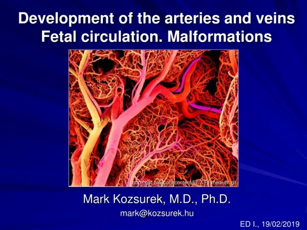
Development of the Veins. Dr. Zeenat Zaidi. The veins develop from the three major venous circuits, vitelline , umbilical and cardinal Like the arteries they develop in a cephalocaudal direction The precursors to the veins are never all present at the same time.

E N D
Development of the Veins Dr. ZeenatZaidi
The veins develop from the three major venous circuits, vitelline, umbilical and cardinal Like the arteries they develop in a cephalocaudal direction The precursors to the veins are never all present at the same time. In addition, as new structures develop the course of veins changes The formation of the liver and the mesonephric kidney has profound affects in redirecting blood flow Most of the venous blood is channeled from the left side to the right side of the body through the anastomosing vessels
Development of the Veins • In a 4 weeks embryo, three paired veins open into the tubular heart: • Vitelline veins, returning deoxygenated blood from the yolk sac • Umbilical veins, bringing oxygenated blood from the placenta. • Common cardinal veins, returning deoxygenated blood from the body of the embryo
Vitelline Veins • Pass through the septum transversum and drain into the sinus venosus • In relation to the liver developing within the septum transversum, the vitelline veins are divided into: • Pre-hapatic part: forms anastomosis around the duodenum which later on gives rise to the portal vein
Hepatic part: interrupted by the liver cords, forms an extensive vascular network called the hepatic sinusoides Post-hepatic part: Left vein disappears Right vein forms the: Hepatic veins & Hepatic segment of inferior vena cava
Umbilical Veins • Bring oxygenated blood from the placenta • Initially run on each side of the developing liver and drain into the sinus venosus • As the liver grows, the umbilical veins loose their connection with heart and open into the liver • The right vein disappears by the end of the embryonic period. The left vein persists
A wide channel, the ductusvenosus, appears through the substance of liver to connect the left umbilical vein with the inferior vena cava • After birth: • The left umbilical vein obliterate to form the ligamentum teres of the liver • The ductus venosus obliterate to form the ligamentum venosum
Cardinal Veins • Are responsible to drain the body of the embryo • The cranial part of the embryo is drained by paired anteriorcardinal veins • The caudal part of the embryo is drained by paired posteriorcardinal veins • The anterior & posterior cardinal veins join to form common cardinal veins, which drain into the sinus venosus
Anterior Cardinal Veins • Become connected by an oblique anastomosis which shunts blood from left to right • This anastomosing channel becomes the left brachiocephalic vein • Left anterior cardinal vein • Cranial part: becomes the left internal jugular vein • Caudal part: degenerates
Right anterior cardinal vein Cranial part: (cranial to the 7thintersegmental vein) becomes the right internal jugular vein Middle part: gives rise to the right brachiocephalic vein Caudal part of rightanterior cardinal vein and the right common cardinal vein form the superior vena cava
Posterior Cardinal Veins • Drain the caudal part of the body of embryo including the developing mesonephroi and largely disappear with this transitory kidneys. • Caudally the two veins get connected by an anastomosing channel that directs the blood from the left to the right vein
Gradually the posterior cardinal veins are replaced by two new veins: subcardinal & supracardinal
The adult derivatives of the posterior cardinal veins are the: Root of the azygosvein & Common iliac veins
Subcardinal Veins • Appear before the supracardinal veins • Become connected: • To each other by the subcardinalanastomosis • With the posterior cardinal veins through the mesonephros • With supracarinal veins through subsupracardinalanastomosis
The adult derivatives of the subcardinal veins are the: Stem of the left renal vein Suprarenal veins Gonadal veins Prerenalsegment of IVC
Supracardinal Veins • Last pair of veins to develop • Appear lateral to the supracardinal veins • Become connected at both ends, to the posterior cardinal veins • Get disrupted in the region of the kidney
Cranial to the kidney: An anastomosis develops between the two veins shunting blood from the left to the right vein Anterior connection of the left vein with the posterior cardinal vein disappears Gives rise to azygos and hemiazygos veins Caudal to the kidney: Leftvein degenerates Rightvein forms postrenalsegmentof IVC
Development of Superior Vena Cava • SVC is derived from the: • Caudal part of the right anterior cardinal vein & • Right common cardinalvein
Development of Azygos Veins • Azygos vein is derived from the: • Cranial part of the right supracardinal vein & • Terminal part of the right posterior cardinalvein • Hemiazygos vein is derived from the cranial part of the left supra-cardinal vein
Development of Inferior Vena Cava • The IVC develops during a series of changes in the primordial veins • Composed of: • Hepatic segmentderived from the right vitellinevein • Prerenal segmentderived from the right subcardinalvein • Renal segment derived from the subcardinal-supracardinalanastomosis • Postrenal segment derived from the right supr-acardinalvein
Anomalies of the Venous System • Persistent left SVC (double SVC): is the most common defect • Left SVC • Absence of IVC • Double IVC
Fetal Circulation & Changes at Birth
Fetal Circulation The main features of the fetal circulation are: • Nonfunctioning lungs • Course of the blood from the placenta to the heart • Three shunts permitting the blood to bypass the liver and lungs: • Foramen ovale • Ductusvenosus • Ductusarteriosus
The oxygenated blood from the placenta reaches the fetus by umbilical vein Most of the blood bypasses the liver through the ductusvenosus, although little blood enters the liver In the inferior vena cava, the oxygenated blood mixes with the deoxygenated blood arriving from the fetus
IVC opens into the right atrium. In the right atrium, the caval blood is guided into the left atrium through the foramen ovale. However, little blood remains in the right atrium, which mixes with the blood arriving through the superior vena cava. In the left atrium also, the oxygenated blood mixes with deoxygenated blood arriving from the lungs.
Blood enters the left ventricle and then into the ascending aorta. Thus the heart and the brain receive better oxygenated blood. The blood from the right atrium enters into the right ventricle, and from there into the pulmonary artery. Most of the blood from the pulmonary artery enters into the aorta through the ductusarteriosus. From the aorta, the blood is distributed to body tissues and flows through the umbilical arteries into the placenta.
The blood circulating in the fetal arterial system is not fully oxygenated There is mixing of oxygenated and deoxygenated blood in the: Liver sinusoids Inferior vena cava Right atrium Left atrium Descending aorta. V IV III II I
What happens at birth? Oh… let me take a deep breath… and then everything will be OK
At birth, dramatic changes occur in the circulatory pattern. • The changes are initiated by baby’s first breath. • Fetal lungs begin to function • Placental circulation ceases • The three shunts that short-circuited the blood during the fetal life ceaseto function
Neonatal Circulation • The infant’s lungs begin to function: • The lungs inflate, which tends to draw blood from the right ventricle. • Oxygenated blood from the lungs passes through pulmonary veins to left atrium. The increased pressure in the left atrium results in closure of the foramen ovale.effectively separating the two atria. • This also increases blood flow to the lungs as blood entering the right atrium can no longer bypass the right ventricle, which pumps it into the pulmonary artery and on to the lungs.
2. The placental circulation ceases. • Umbilical vessels are no longer needed. They become obliterated • Occlusion of the placental circulation causes fall of blood pressure in the inferior vena cava and right atrium 3. The shunts stop to function: • Within a day or two of birth, the ductusarteriosus closes off, preventing blood from the aorta from entering the pulmonary artery • The ductusvenosus closes off so that all blood entering the liver passes through the hepatic sinusoids.
Adult Derivatives of Fetal Vascular Structures • Ductusvenosusbecomes the ligamentumvenosum, attached to the inferior vena cava. • Ductusarterioususbecomes ligamentumarteriousum • Foramen ovalecloses shortly after birth, fuses completely in first year and becomes fossaovalis • The intra-abdominal portions of the umbilical arteries become the medial umbilical ligaments • The intra-abdominal portion of the umbilical vein becomes the ligamentumteres. (the umbilical vein remainspatent for a long time and may be used for exchange transfusions during early infancy. The lumen of the umbilical vein usually does not disappear completely; hence, the ligamentumteres can be cannulated in adults for the injection of contrast medium or chemotherapeutic drugs).
Persistence of Fetal Circulation • Patent ductus arteriosus and patent foramen ovale each characterize about 8% of congenital heart defects. • Both cause a mixing of oxygen-rich and oxygen-poor blood • Blood reaching tissues is not fully oxygenated and can cause cyanosis. • Many of these defects go undetected until child is at least school age.