Download
1 / 49
660 likes | 2.3k Views
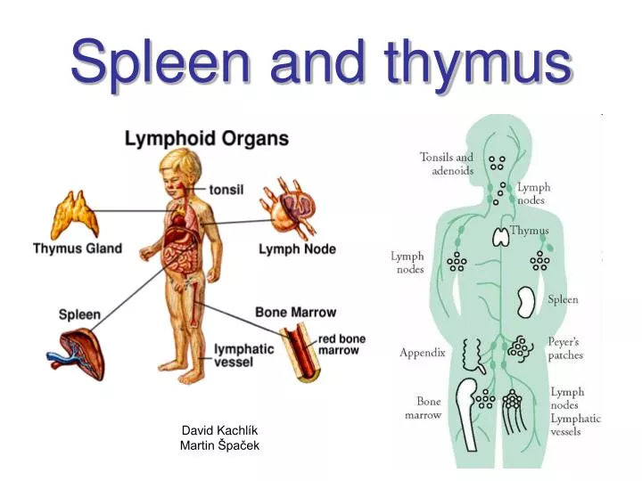
Spleen and thymus. David Kachlík Martin Špaček. Lymphoid organs. Primary (central) lymphoid organs Thymus Bone marrow = Medulla ossium Secondary (peripheral) lymphoid organs Spleen = Splen (lien) Lymph nodes = Nodi lymphoidei Tonsils = Tonsillae Lymph nodules lymphoidei.

E N D
Spleen and thymus David Kachlík Martin Špaček
Lymphoid organs • Primary (central) lymphoid organs • Thymus • Bone marrow = Medulla ossium • Secondary (peripheral) lymphoid organs • Spleen = Splen (lien) • Lymph nodes = Nodi lymphoidei • Tonsils = Tonsillae • Lymph nodules lymphoidei
Thymus - history Jacques Miller • 2nd cent. - Galen: „organ of mystery“ • 19th cent. • fills space between the lungs • blamed for sudden death infant syndrome („thymic asthma“; „status thymolymphaticus“) • 1905 - first „successful treatment“ by radiotherapy • 1961 - discovery of the function of the thymus by Jacques Miller • 1974 - John Caffey: • „most mistakes I’ve seen were not because one didn’t know some disease, but because he didn’t know he was looking at normal.“
Thymus • lymphoepithelial organ • primary lymphoid organ • lobus dx. et sin. • lobuli, cortex, medulla • (lobuli thymici accessorii) • relatively largest after birth (12-14g) • begins to atrophy in puberty • remnants still evident in older age
mediastinum superius behind sternum 20-50 gnewborn 16 g (10-35 g)from below thyroid gland down to pericardium • successive atrophy from puberty • replaced with adipose tissue after 50th year of age (5-15 g)
Thymus • mediastinum superius (1st layer) • covered with mediastinal connective tissue
case-report age: 2 month dg: coarctation of aorta
Thymus – blood vessels • branches from: • a. thyroidea inf. • thoracica int. (a. pericardiacophrenica) • arcus aortae • non-fenestrated capillaries • haematothymic barrier = claustrum haematothymicum • cortex • endothelium of capillaries • basal lamina of capillaries (+ pericytes, resp.) • connective tissue layer (+ macrophages) • basal lamina of reticular cells • reticular cells
Thymus - development • ventral process of 3rd (and 4th) pharyngeal pouch • mediocaudal descent (4th-7th week) • endoderm proliferation • 10th week:colonization with stem cells (lymphocytes) from blood islets, liver and bone marrow • connective tissue septa ingrow from mesenchyme
Thymus - structure • covered with mediastinal connective tissue • contains vessels • grows into thymic tissue – false lobules • septa corticalia lobuli thymici • cortex thymi • darker apperance • medulla thymi • lighter apperance
Thymus - cortex • reticular epithelium (cytoreticulum corticale) • epitheliocyti reticulares (stellate cells connected with desmosomes) – typy I-III • they form a spatial network • macrophages a thymocytes nurse cells • thymocytes (small and frequent) • mainly T-lymfocytes • rapidly multiply during development • cellula thymocytopoietica progenetrix prothymocytus thymocytus corticalis
HE (x 480) immunoperoxidase (x 100) Thymus - cortex
Thymus - medulla • reticular epithelium (cytoreticulum medullare) • type IV-VI • thymocyti medullares (small and middle) • not so densely • corpuscula thymica (Hassal‘s bodies) • 30-150 μm • concentric layers of flattened reticular cells • keratinization, karyolysis • arise and fade out repetitively • noduli lymphoidei thymici • dendritic cells • antigen-presenting cells (APC) • myoid cells • function not clear, possible role in pathogenesis of myastenia gravis?
Thymus - histophysiology • multiplication of T-lymfocytes • they leave thymusvia medulla (10 %) • travel to lymph nodes, Peyer‘s patches, spleen • many growth factors proliferation and diferentiation of T-lymfocytes • corticoids and sexual hormones supress proliferation and accelerate involution
Thymus - developmental disroders • aplasia thymi • aplasia thymoparathyroidea (DiGeorge‘s syndrome) • few B-, no T-lymfocytes • ectopia thymi • hypoplasia thymi (Sprintzen‘s syndrome) • textus thymicus accessorius intrapericardial accessory thymic tissue situated ventral to aorta ascendens
DiGeorge‘s syndromeAplasia thymoparathyroidea syndrome of 22q11.2 deletion 1:3000
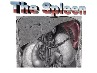
Spleen(splen, lien) • tunica serosa (peritoneum) • capsula (tunica fibrosa) • facies • diaphragmatica • visceralis (renalis, gastrica, colica, pancreatica) • extremitas anterior + posterior • margo inferior + superior (obsolete “margo crenatus“) • hilum splenicum (medially) • sinus splenicus
SPLEEN view from right anterior
Spleen (splen, lien) • length 10-13 cm; width 6-8 cm; thickness 4 cm • weightdepends on the blood fillling • ♂ 140-160 g / ♀120-150 g • weight 200 g is not pathological • lig. splenorenale, gastrosplenicum, splenocolicum, phrenicosplenicum • splen accessorius (= spleniculus) • 4-6 segments
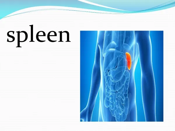
Spleen – position and syntopy • intraperitoneal organ • left hypochondrium • 4 cm lateral to medioclavicular line • 9th-11th rib, lonf axis along 10th rib • normally not palpable • bursa omentalis • cavitas pleuralis
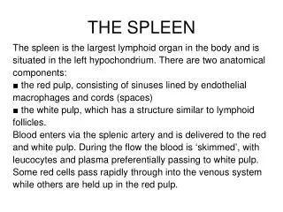
Spleen – structure • fibrous capsule (collagene) • sparse smooth muscle cells • covered with serosa (except hilum) • fibrous septa into pulp (trabeculae splenicae) • pulpa splenica • pulpa alba • zona marginalis • noduli lymphoidei splenici • pulpa rubra • Billroth‘s cords • reticular connective tissue
Spleen - characteristic • secondary lymphoid organ • largest lymphoid organ • imunologic blood filter • removal of microorganisms • „cemetery“ of erythrocytes • storage of blood • hematopoiesis during development
Spleen – blood supply • truncus coeliacus a. splenica rr. splenici aa. trabeculares arteriolae vaginatae pulpae albae • within periarterial lymphatic sheath (PALS; vagina lymphoidea periarteriolaris) • arteriolae centrales (nodulares) within noduli lymphoidei splenici • sinuses of zona marginalis • aa. pulpae rubrae aa. penicillares arteriolae penicillares • vagina perioarteriolaris macrophagocytica (Schweigger-Seidel‘s caspule) • vasa sinusoidea splenica (in pulpa rubra) • open x closed circulation • fusiform endothelial cells, clefts, interrupted basal lamina vv. pulpae rubrae vv. trabeculares v. splenica v. portae
Spleen – white pulp • reticular connective tissue with lymphocytes • cords ensheath arteries– PALS (vagina lymphoidea periartriolaris) • T-lymphocytes • arteries within nodules located excentriclly • Malpighi‘s corpuscle • B-lymphocytes • zona marginalis – between pulpa rubra/alba • sinuses and lymphoid tissue • macrophages (antigen presentation)
Spleen – red pulp • Billroth‘s cords (chordae splenicae) • cells between sinusoids • lymphocytes, macrophages, erythrocytes • reticular fibers (fibrae reticulares anulares) – hoop arrangement • blood sinuses • fusiform endothelial cells (endotheliocyti fusiformes), interrupted (endothelium disjunctum) • located close to reticular fibers • spatium intersinusoideum splenicum
Spleen - histophysiology • immune function of lymphocytes • destruction of erythrocytes
Spleen – clinical relevance • splenomegaly (viral mononucleosis, liver disease, blood cancers - lymphoma and leukemia) • hypersplenism • „two-stroke“ rupture of spleen • abdominal injury • splenectomy • higher tendency to encapsulated bacteria (pneumococcus, meningococcus, haemophilus) • mandatory vaccination • sickle-cell disease • thrombocytopenia
Spleen • X-ray • US
Spleen - development • derived from mesenchyme of dorsal mesogastrium (mesenchyma splanchnopleurale) • from 5th week • mesenchymal cells – diferentiation • capsule • reticular net • parenchyma • 4th month - hematopoiesis • from 2nd month - lymhopoiesis
Spleen – developmental disorders • asplenia • conjunction splenogonadalis, splenopancreatica • lobulatio splenis • nodulus splenicus accessorius • polysplenia • splen accessorius (gl. suprarenalis, pancreas, gaster, intestinum) – 10 % • splen migrans
Splenaccessorius 1 – hilum splenicum 2 – pancreas (or its vicinity) 3 – lig. splenocolicum 4 – omentum majus 5 – mesenterium 6 – apertura pelvis superior 7 – ovarium/tuba uterina 8 – scrotum 9 – hiatus oesophageus diaphragmatis 10 – omentum minus