Download
1 / 25
470 likes | 1.38k Views
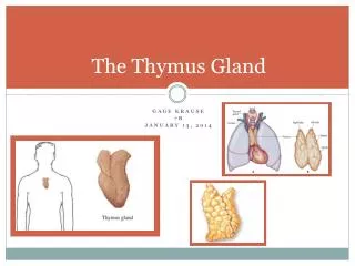
Thymus. Thymus. Thymus. Primary lymphatic organ Anterior mediastinum Present – Infants – larger Adults – degenerated & infiltrated by adipose tissue. Thymus. Covered – Capsule - Dense irregular connective tissue Collagen fiber Elastic fibers with fibroblast

E N D
Thymus • Primary lymphatic organ • Anterior mediastinum • Present – • Infants – larger • Adults – degenerated & infiltrated by adipose tissue
Thymus Covered – Capsule - • Dense irregular connective tissue • Collagen fiber • Elastic fibers with fibroblast • Septa (trabecula) • incomplete lobules
Thymus Parenchyma • Epithelioreticular cells • Has lobules • Outer cortex • Inner medulla
Thymus • Cortex • Thymocytes, • Macrophages, • Dispersed reticulo - epithelial cells.
Medulla Stains lightly because lymphocytes are not densely packed Hassall’s corpuscle / thymic corpuscle
Hassall’s corpuscle / thymic corpuscle • They are situated in the medulla of thymus. • It has a central core formed by degenerated epithelial cells surrounded by concentrically arranged epithelial cells. • These cells form a pink staining hyaline mass.
Thymus Blood vessel Thymic lobule Interlobular septum Cortex Medulla Hassall’s corpuscle
Hassall’s corpuscle Thymic lobule Medulla Cortex
Blood thymus barrier • Blood vessels entering the thymus from the trabeculae get surrounded by the • sheath of connective tissue, • epitheliocyte
Blood thymus barrier • Prevent most circulating antigens from leaving vasculature and entering the thymic cortex.
Functions Of Thymus • Thymus act as a central organ of lymphatic system • It is essential in early weeks of neonatal life and regulates the growth of peripheral lymphoid tissue. • Thymus is enlarged in some autoimmune diseases(myasthenia gravis).
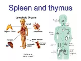
PALATINE TONSIL • Present – oropharynx • Called diffuse lymphatic tissue Because lymphoid follicles – present just beneath lining epithelium.
PALATINE TONSIL • Lined – stratified squamous non keratinized epithelium • Tonsillar crypts - epithelium dips – substance of tonsil • Lymphatic nodules with germinal center – present just beneath epithelium • There are • B & T lymphocytes • Mucous acini are seen
Palatine tonsil Stratified squamous non-keratinized epithelium Lymphoid follicle Tonsillar crypt Mucous acini
Palatine tonsil- magnified Stratified squamous non-keratinized epithelium Tonsillar crypt Mucous acini Lymphoid follicle