Download
1 / 24
260 likes | 1.19k Views
Jugular venous pressure and carotid arterial pressure. Maha Alenazy. Objectives. What are Jugular venous pressure and Carotid arterial pressure their normal recording Abnormalities and clinical importance Difference between JVP and CAP JVP, CAP, and ECG. Jugular Venous Pressure.

E N D
Jugular venous pressure and carotid arterial pressure MahaAlenazy
Objectives • What are Jugular venous pressure and Carotid arterial pressure • their normal recording • Abnormalities and clinical importance • Difference between JVP and CAP • JVP, CAP, and ECG
Jugular Venous Pressure • The jugular venous pressure (JVP) provides an indirect measure of central venous pressure. • The internal jugular vein connects to the right atrium without any intervening valves - thus acting as a column for the blood in the right atrium. • The JVP consists of certain waveforms and abnormalities of these can help diagnose certain conditions.
How to examine • Use the right internal jugular vein (IJV). • Patient should be at a 45° angle. • Head turned slightly to the left. • If possible have a tangential light source that shines obliquely from the left. • Locate the surface markings of the IJV - runs from medial end of clavicle to the ear lobe under medial aspect of the sternocleidomastoid. • Locate the JVP - look for the double waveform pulsation (palpating the contralateral carotid pulse will help). • Measure the level of the JVP by measuring the vertical distance between the sternal angle and the top of the JVP. Measure the height - usually less than 3 cm.
JVP • Classically three upward deflections and two downward deflections have been described. • The upward deflections are the "a" (atrial contraction), "c" (ventricular contraction and resulting bulging of tricuspid into the right atrium during isovolumic systole) and "v" = atrial venous filling. • The downward deflections of the wave are the "x" (the atrium relaxes and the tricuspid valve moves downward) and the "y" descent (filling of ventricle after tricuspid opening). • Jugular Venous Pulse
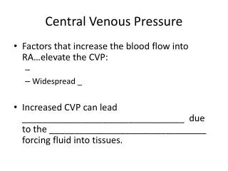
Abnormal JVP Causes of a raised JVP: • Heart failure • Constrictive pericarditis (JVP increases on inspiration called Kussmaul's sign) • Cardiac tamponade • Fluid overload e.g. renal disease • Superior vena cava obstruction (no pulsation)
Abnormalities of the a wave • Disappears in atrial fibrillation • Large a waves occur in any cause of right atrial contraction pressure (pulmonary hypertension and pulmonary stenosis) and tricuspid stenosis, right heart faluire. • Extra large a waves (called cannon waves) in complete heart block, atrial flutter, and tachycardia • Prominent v waves • Tricuspid regurgitation - called cv or V waves and occur at the same time as systole (combination of v wave and loss of x descent); there may be ear lobe movement
Cont. • Slow y descent • Tricuspid stenosis • Right atrial myxoma • Steep y descent • Right ventricular failure • Constrictive pericarditis • Tricuspid regurgitation • (The last two conditions have a rapid rise and fall of the JVP called Friedreich's sign)
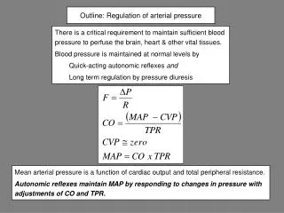
Carotid arterial pressure • The carotid pulse is a pulse that can be taken on the right side of the neck over the carotid artery in order to determine heart rate • It is considered to be a more reliable site to measure than the wrist, particularly in individuals who have suffered some kind of trauma and/or who are in shock
Cont. • Normally, the carotid pulse can be felt by the fingers, but in some conditions, such as shock and low blood pressure, an instrument called a doppler may be needed to find the carotid pulse and amplify it so it can be heard. Conditions that can afflict the carotid pulse include arterial clots, arterial embolisms, hemorrhage, shock, cardiac arrhythmias, and cardiac arrest.
CAP RECORDING • A tracing of the normal carotid pulse involves a smooth, rapid upstroke with a dome-shaped peak
How to examine • examined with the patient supine and the trunk of the patient's body slightly elevated • The fingers should be positioned between the larynx and the anterior border of the sternocleidomastoid muscle at the level of the cricoid cartilage • In palpating the pulse, the degree of pressure applied to the artery should be varied until the maximum pulsation is appreciated.
Cont. • Palpation of the carotid artery normally detects a smooth, fairly rapid outward movement beginning shortly after the first heart sound and cardiac apical impulse • The pulse peaks about one-third of the way through systole • This peak is sustained momentarily and is followed by a downstroke that is somewhat less rapid than the upstroke
CAP and ECG The ascending limb is called the anacrotic limb. It is due to the rapid ejection of the left ventricle to the aorta (pressure up in aorta). (anacrotic limb is due to stroke volume and artery compliance. The descending limb (diacrotic limb) is due peripheral resistance as blood continues to flow to the peripheral arteries. When blood is in aorta and at end of systole some blood tries to go back closing aortic valve. This creates some elevation in the pressure causing: diacrotic notch or (Incisura)
Abnormal CAP • The dicrotic pulse has two palpable waves, one in the systole and one in diastole (Figure Below). It usually denotes a very low stroke volume, particularly in patients with dilated cardiomyopathy.
Cont. • A dicrotic pulse results from an accentuated dicrotic wave and tends to occur in patients with*sepsis, *severe heart failure, *hypovolemic shock, *cardiac tamponade, and *aortic valve replacement.
Cont. • Pulsusbisferiens with both percussion and tidal waves occurring during systole. • observed in patients with hemodynamically significant aortic regurgitation or combined aortic stenosis and regurgitation with dominant regurgitation.
Cont. • the pulse wave upstroke rises rapidly and forcefully, producing the first systolic peak ("percussion wave"). A brief decline in pressure follows because of the sudden midsystolic decrease in the rate of left ventricular ejection, when severe obstruction often develops. This pressure trough is followed by a smaller and more slowly rising positive pulse wave ("tidal wave") produced by continued ventricular ejection and by reflected waves from the periphery.
Cont. Abnormal • Carotid pulses: • A, hyperkinetic pulse; • B, bifid pulse; • C, bifid pulse characteristic of IHSS; • D, hypokinetic pulse; • E, pulsusparvus et tardus
JVP vs. CAP The JVP pulse is • Not palpable • Occludable: Obliterated by pressure • Characterised by two beats per cardiac cycle. • Varies with respiration - decreases with inspiration • Varies with Head-Up Tilt (HUT). Stand up will lower it.
JVP, CAP, ECG a after the P wave while c after ORS A, c with S1 and x ends before S2 while v is a bit after S2.
Some Tips! • In a normal person,the a wave is larger than v wave and x descent is more prominent than y descent • A wave occurs just before S1or carotid pulse and has a sharp rise and fall • V wave occurs just after arterial pulse and has a slow undulating pattern • X descent occurs between S1 and S2 and y descent well after S2