Download
1 / 53
1.32k likes | 5.22k Views
Jugular Venous Pulse. Dr .Latchumanadhas Madras medical mission. History of JVP. Lancis : Venous pulse of EJV Chauvea,marey :Graphic recording of JVP Pontain :Wave pattern in JVP James Mackenzie :Nomenclature of JVP

E N D
Jugular Venous Pulse Dr .Latchumanadhas Madras medical mission
History of JVP • Lancis : Venous pulse of EJV • Chauvea,marey :Graphic recording of JVP • Pontain :Wave pattern in JVP • James Mackenzie :Nomenclature of JVP • Paul wood : Hemodynamics of JVP
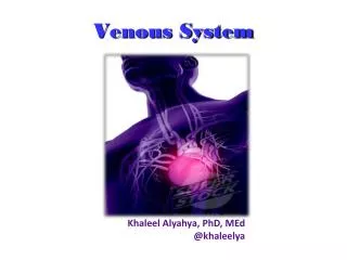
Jugular Venous System • Venous system contains 70-80% of the circulating blood volumes . • Right atrial and right ventricular filling produce pulsations in the central veins that are transmitted to jugular veins . • An accurate assessment of the jugular venous pulse reflects the hemodynamics of the right sided heart . • Right atrial pressure during systole and right ventricular filling pressure during diastole are producing pulsations and pressure waves in jugular veins .
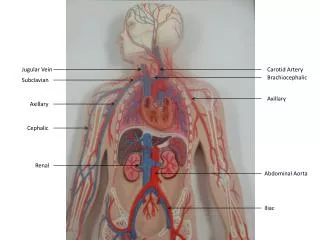
Exam of JVP • Right IJV is usually assessed both for waveform and estimation of CVP • Internal jugular venous pulsation located between two heads of sternocleidomastoid muscle are transmitted to overlying skin . • Right IJV is in direct continuity with SVC and right atrium • Left IJV drains into left innominate vein, which is not in straight line from RA • Right IJV and innominate vein is not compressed by adjacent structures
Right IJV Preferred :Why? • Direct continuation of right atrium • Straight line course through innominate vein to the svc and right atrium • IJV is less likely affected by extrinsic compression from other structures in neck • There are no or less numbers of valves in IJV than EJV • Less impact of vasoconstriction on IJV due to sympathetic activity than EJV
Superficial and lateral in the neck Better seen than felt Has two peaks and two troughs Descents >obvious than crests Digital compression abolishes venous pulse Jugular venous pressure falls during inspiration Abdominal compression elevates jugular pressure Mean jugular venous pressure falls during standing Deeper and medial in the neck Better felt than seen Has single upstroke only Upstroke brisker and visible Digital compression has no effect Do not change with respiration Abdominal compression has no effect on carotid pulse Carotid pulse do not change when standing . Differences between IJV and Carotid pulses
Position of Patient • Patient should lie comfortably and trunk is inclined by 45 degree position • Elevate chin and slightly rotate head to the left • Inclination angle should be subtended between trunk and bed , while neck and trunk should be in same line • When neck muscles are relaxed ,shining the light tangentially over the skin and see pulsations • In patients with low jugular pressure , a lesser (<30‘) inclination is desirable • In patients with high jugular pressure ,a greater (60-90‘) inclination is required to obtain visible pulsations • Simultaneous palpation of the left carotid artery and or apical impulse aids in timing of the venous pulsations in cardiac cycle .
Normal JVP • Normal JVP reflects phasic pressure changes in RA during systole and RV during diastole • Two visible positive waves ( a and v) and two negative troughs ( x and y) • Two additional positive waves can be recorded . C wave interrupts x descent and h wave precedes the next a wave
Normal JVP Waveform • Consists of 3 positive waves • a,c & v • And 3 descents • x, x'(x prime) and y
a Wave • First positive presystolic a wave is due to right atrial contraction results in retrograde blood flow in to svc and jugulars • Effective RA contraction is needed for visible a wave • Dominant wave in JVP and larger than v • It precedes upstroke of the carotid pulse and S1, but follow the P wave in ECG
x Descent • Systolic x descent (systolic collapse) is due to atrial relaxation during atrial diastole • X descent is most prominent motion of normal JVP which begins during systole and ends just before S2 • It is larger than y descent • X descent more prominent during inspiration
c WAVE • Second positive wave recorded in JVP which interrupts the x descent • Produced by carotid artery impact on JVP upward bulging of closed TV into RA during isovolumic contraction
x` Descent • x`descent is systolic trough after c wave • Due to fall of right atrial pressure during early RV systole downward pulling of the TV by contracting right ventricle descent of RA floor by contracting RV
v Wave • Third positive wave in JVP which begins in late systole and ends in early diastole • Rise in RA pressure due to continued RA filling during ventricular systole when tricuspid valve closed • It is roughly synchronous with carotid upstroke and corresponds S2 .
y Descent • Diastolic collapse wave (down slope v wave) • It begins and ends during diastole well after S2 • Decline of RA pressure due to RA emptying during early diastole when tricuspid valve opens • Initial y descent is corresponds to rapid RV filling and later part of y wave is produced by continued diastolic inflow in to RV .
h wave • Small brief positive wave following y descent just prior to a wave during period of diastasis • Described by Hieschfelder in 1907 • It usually seen when diastole is long (as in slow heart rates) • With increasing heart rate, y descent immediately followed by next a wave .
Prominent a Wave • Forceful atrial contraction when there is resistance to RA emptying or increased resistance to ventricular filling • RV inflow obstruction: Tricuspid stenosis or atresia RA mxyoma Decreased ventricular compliance: Pulmonary stenosis Pulmonary hypertension of any cause RV infarction RV cardiomyopathy (including HOCM) Acute pulmonary embolism
Cannon Waves • Whenever RA contracts against closed TV valve during RV systole : Systolic cannon wave . • Venous corrigan by paul wood • Regular cannon waves: Junctional rhythm VT with 1:1 retrograde conduction Isorhythmic AV dissociation • Irregular cannon waves : Complete heart block Classic AV dissociation Ventricular pacing or ventricular ectopics .
Absent a Wave • When no effective atrial contraction as in atrial fibrillation • In atrial fibrillation , both atrial contraction as well as relaxation become in effective • In sinus tachycardia , when a wave may fuse with preceding v wave , especially when the PR interval is prolonged .
Abnormal x descent • Prominent x descent : Presence of atrial relaxation with intact tricuspid valve and good RV contraction is needed for prominent x descent. • Causes : Constrictive pericarditis Atrial septal defect
Absent x descent • Moderate to severe TR: EARLY SIGN • Obliteration of x descent with early build up of large systolic wave ( cv wave) • RV dysfunction due to PHT with RV failure severe RV infarction • Chronic atrial fibrrilation : loss of atrial relaxation
Prominent v wave • Increased RA volume during ventricular systole produce prominent v wave • Severe TR : giant v wave (Lanci sign) – ventricularization of atrial / jugular pressure • Giant v wave sometimes causes : systolic movement of ear lobe head bobbing with each systole systolic pulsation of liver pulsatile exophthalmos pistol shots heard over IJV
PROMINENT V WAVE • Tricuspid regurgitation • ASD with mitral regurgitation • VSD of LV to RA shunt (Gerbode's defect) • Severe congestive heart failure • Cor pumonale • Atrial fibrillation
Rapid y Descent • Severe TR • C .Pericarditis (Friedreich's sign): Early rapid ventricular filling • PHT with RV dysfunction • Severe RV failure • ASD with mitral regurgitation • Severe RV infarction
Slow y Descent • When RA emptying and RV filling are impaired , y descent is slow and gradual Tricuspid stenosis Right atrial myxoma Pericardial tamponade( y descent may even be absent).
DD of Double Descents • Costrictive pericarditis • RV infarction with mild RV dysfunction • PHT with early RV decompensation DD of single Descent x descent only : cardiac tamponade y descent only : severe TR with RV dysfunction chronic AF with CCF EMF with TR
Estimation of Venous Pressure • Measuring jugular venous pressure • Hepatojugular reflux • Examining the veins on the dorsum of the hand • Examining the veins of the undersurface of the tongue • Assessment of jugular venous pressure at bed side reflect mean right atrial pressure
Measurement of JVP • Sternal angle or angle of Louis is a surface anatomical mark ,reference point used for JVP measurement (Paul wood) • It is found approximately 5 cm above the center of the right atrium • Distance between sternal angle and center of right atrium remains relatively constant regardless of position of the thorax • Use sternal angle as zero reference point • Examine the patient in an optimal position(45`recline) and relaxed neck muscles , shining the light tangentially across the skin overlying IJV and see the top of oscillating venous column
Measurement of JVP • Two scale method is commonly used • A horizontal scale at the top of the oscillating venous column in IJV cuts the vertical scale at the sternal angle gives JV pressure in cm of water • Normally JV pressure does not exceed 4 cm above the sternal angle • Since RA is approximately 5 cm below the sternal angle , the jugular venous pressure (RA mean pressure) is corresponds to 9 cm • By way of conversion , normal mean JV pressure does not exceed 7 mm Hg (9 cm column of water / 1.3 =6.9) • Elevated JVP : JVP of >4 cm above sternal angle .
Elevated JVP • Increased RVP and reduced compliance: Pulmonary stenosis Pulmonary hypertension Right ventricular failure RV infarction • RV inflow impedance: Tricuspid stenosis / atresia RA myxoma Constrictive pericarditis
Elevated JVP • Circulatory overload : Renal failure Cirrhosis liver Excessive fluid administration • SVC obstruction • COPD
Kussmual's sign • Mean jugular venous pressure increases during inspiration as a result of impaired RV compliance. • Constrictive pericarditis • Severe right heart failure • RV infarction • Restrictive cardiomyopathy
Abdominal -Jugular Reflux • Hepatojugular reflux – Rondot (1898) • Apply firm pressure to periumbilical region for 10 -30 sec with patient lying comfortably and breathing quietly while observe JVP • Normally JV pressure rises transiently(15 sec) to < 3cm while abdominal pressure is continued • If JV pressure remains elevated until abdominal pressure is continued: called as Positive AJR.
Positive AJR • Incipient and or compensated RVF • LVF with volume overload • Tricuspid regurgitation • False Positive AJR ( without CCF) COPD Systemic vasoconstriction Increased sympathetic tone Severe anaemia
Gaertner's method • Measurement of JVP by examining the veins on the dorsum of the hand • When patient sitting or lying at a 45‘ elevation , arm slowly and passively raised from dependant position until the vein collapses • Height of the limb above the level of sternal angle at which vein collapses represents the venous pressure • When venous pressure is normal , veins of hand collapse at the level of sternal angle
Cardiac tamponade • JVP is usually elevated • a wave never prominent • v and x wave is normal • y descent is diminished or absent • Kussmaul's sign usually negative
Constrictive pericarditis • JVP is elevated • a wave is usually normal • v wave is usually equal to a wave • x descent –prominent • y descent – rapid descent • Kussmauls sign is usually positive
Endomyocardial fibrosis • JVP is usually elevated • a wave is prominent • v wave is prominent due to TR • x descent is obliterated due to TR • Rapid y descent is due to TR • Kussmaul's sign is negative
Primary Pulmonary Hypertension • Normal RV compliance :normal JVP • Early RV decompensation : JVP may be elevated a wave is prominent and larger than v wave x and y descent seen and equal Decompensated RVF: JVP is always elevated a and v wave prominent , v wave larger than a wave x descent is diminished or absent rapid y descent due to TR
JVP in ASD • JVP is normal and equal a and v waves. x descent is more prominent . • Elevated JVP may seen in severe PAH and in RVF • Prominent a wave with PS and MS • Prominent v wave with PAH and in RVF with TR • Rapid y descent with RVF or TR
JVP in VSD • Prominent a wave with severe PS • Elevated JVP with CHF • Prominent v wave with Gerbode's shunt • In Eisenmenger complex : JVPressure usually normal Normal a and v waves CHF and TR is rare
Ebstein Anomaly • JVP is usually normal • Prominent a waves are seen only occasionally • Attenuated x descent and systolic v wave are not reflected in jugular pulse despite appreciable TR • Unimpressive JVP is attributed to damping effect of large capacitance RA and thin, toneless atrialized RV (Hypokinetic TR) • Prominent a and v / elevated JVP with advent of right ventricular failure .
Cyanosis with prominent a wave • It usually indicates intact IVS • Severe PS with intact IVS and Right to Left shunt • Tricuspid atresia • TAPVC with restrictive ASD • Eisenmenger syndrome with PDA • PPH with right to left shunt via PFO