Download
1 / 59
610 likes | 977 Views
Identification of people alive , cadavers and human remains. Agata Thannhauser.

E N D
Identification of peoplealive, cadavers and humanremains Agata Thannhauser
Traffic accidents, natural disasters and crime entail numerous fatal victims, whose identity is often unknown. Depending on the circumstances of finding the bodies the particular methods are used to identify. These methods differ in degree of credibility. Thelist of identification techniques of dead bodies was put forward at the 5th Interpol’s Meeting on the identification of victims of mass disasters and natural disasters, held in 1993 at Interpol’s headquarters in Lyon.
Problems • Physical appearance - short-term changes in facial expression occurring while blinking, speaking, expressing emotion or communicating. Also important are slow changes in the appearance caused by aging or changing image
The geometry of the acquisition - usually the face in the picture is in an unknown location, it is not known which way it is facing and it is not known what is the size (scale)
Conditions of imaging - human face lighting can have a big impact on the appearance on the photo
Distortion of compression - they are often quite unexpected worsening of image quality caused by the compression (in order to send or archive)
Identificationof personal items, jewelry, etc • Final places on the list of techniques of body identification, as presented at the 5th Interpol’s Meeting, belong to identification of personal items, jewelry, etc, identification on the basis of identity documents, and identification by witnesses, family members or friends. • Such a low degree of reliability attributed to personal items and documents of identity items from the fact that, in forensics, such things have been proven to easily change owners, either intentionally or accidentally. • A low degree of reliability is also attributed to identification by family members or friends. The reason for this is making common, unintentional mistakes in identifying the dead body due to stress or corpse condition which prevent recognition even by closest relatives.
Identificationof personal items, jewelry, etc One of the methods used to identify is the identification based on things found on or next to the remains. However it is necessary to remember , that this is only an auxiliary method –we can never confirm the identification based on this method. In the case of unidentified human corpses we describe the clothing very accurately (including size, color, texture of the material) shoes - including size, brand name, individual characteristics like tailor's alterations.
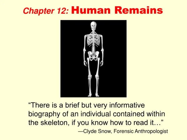
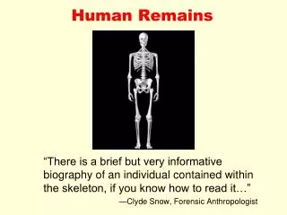
ACCURACY OF SEXING….KROGMAN • ENTIRE SKELETON… 100% • PELVIS & SKULL…….98% • PELVIS………………..95% • SKULL…………………90% • LONG BONES………..80%
Determining the Sex of Unknown Human Skeletal Remains Male pubis (ventral) arrows show locationwhereventralarcwould be located, thisfeatureisabsentinmales Femalepubis (ventralaspect): arrowsindicatetheventralarc VENTRAL ARC
Determining the Sex of Unknown Human Skeletal Remains Femalepubis (dorsalview): bracketshowingarea of ischiopubicconcavity Male pubis (dorsal) bracketindicatingabsence of concavity
Determining the Sex of Unknown Human Skeletal Remains Male sacroauricular joint. Bracketedareademonstratingabsence of pre-auricularsulcus. Femalesacroauricular joint withpre-auricularsulcus. Arrowsshowingpitsinthesulcus
Determining the Sex of Unknown Human Skeletal Remains Male nuchalcrestwith an inionhook Femalenuchal region – notetheabsence of rugosity
Determining the Sex of Unknown Human Skeletal Remains Male mastoid Femalemastoid
Determining the Sex of Unknown Human Skeletal Remains Differenceinthefrontal Slantedwithlargebrowridgesinmales High and roundedwithoutbrowridgesinfemales
Other bones are not usually as good an indicator regarding sex
SUMMARY • Becausemalesgenerallyarelarger and morerobustthanfemales, theskeletons of thesexescan be distinguished by simplyobservingsize and rugosity. • Theare a number of a shapecharacteristics of thefemalepelvis, related to childbirth, thatdifferntiateitfromthepelvis of males. • Theishium-pubicindex, whichquantifiesthelongerpubicbone of femaleswhencompared to males, istheonlycommonlyusedmetricmethod for distinguishingthesexes. • Theskulls of malesgenerallyarelarger and morerugged, withlargemastoidprocesses and browridges, thantheskull of females. • A number of metricmeasurements of theskullcan be usedwithdiscriminantfunctions to differentiatethesexes. • A number of measurements of thepostcranialbonescan be used to separatemalesfromfemales.
Determination of agefrombones • Ages 0-5: teeth are best – forensic odontology • Ages 6-25: epiphyseal fusion – fusion of bone ends to bone shaft • epiphyseal fusion varies with sex and is typically complete by age 25 • Ages 25-40: very hard, can use pubic symphysis • Ages 40+: periodontal disease, arthritis, breakdown of pelvis, occupational stress, unique clues
AGE ESTIMATION METHOD Fusionstates of cranialsutures. . The use of cranial suture closure for estimating age has a long and controversial history. In the mid1920s, T. Wingate Todd and D. W. Lyon published a series of articles describing changes in the cranial vault sutures related to age .They discovered that these lines were open in young people but had a tendency to close through time until, by old age, they were completely obliterated. Despite this promising start, their research was flawed because they discarded specimens from their study that exhibited anomalous closures. Over 13% of Whites and 34% of Blacks that were originally part of their sample wereexcluded during analysis. Although these attempts to choose only specimens that fit a preconceived notion of "normal" were well intentioned, modem scientific theory renders this unacceptable. In addition, even though they culled their sample to obtain more homogeneous results, Todd and Lyon still encountered a high amount of variability, which made age estimates based on their method prone to considerable error. Thus, as time passed, many forensic (and other) anthropologists simply did not use this method CRANIAL AND PALATE SUTURES
AGE ESTIMATION METHOD Fusionstates of cranialsutures. open beginningfusion Buikstra and Ubelaker (1994) recognize four stages of closure: open, mini-mal, significant, and obliteration. Open refers to sutures where there is no evidence of closure .The gap between the bones is easily discernable and appears as a "bottomless" groove separating the bones.- Minimal is characterized by the closing of these gaps by bridges of bone that vary from a single connection to bridges that encompass less than 50% of the entire suture . CRANIAL AND PALATE SUTURES
AGE ESTIMATION METHOD Fusionstates of cranialsutures. Finally, obliteration refers to complete fusion between bones with no discernible gap; the sutures either appear as lines drawn on the bones or are no longer visible Significant closure means that there is more than 50% fusion between the bones, but with some parts still open. significantfusion obliteration CRANIAL AND PALATE SUTURES
AGE ESTIMATION METHOD PUBIC SYMPHYSIS • In his pioneering article, T. Wingate Todd (1920) described the alterations that occur in the pubic symphysis over time and how these can be used to estimate age at death. Todd described 10 stages of changes to this structure that, occur during aging, with time ranges of uneven lengths (e.g., 18 to 19, 30 to 35, 50+) for each stage. Since his initial research, the value of using pubic face changes to evaluate age at death has received much attention. In 1955, Sheila Brooks, feeling that the age ranges for each stage were too high, modified Todd's time ranges down by several years in each category. Although the schedules and stages vary, all the abovementioned techniques look at the same features originally described by Todd: • the bone of the pubic face, • the ventral and dorsal margins, • the upper and lower extremities, • ossific nodules
The bone of the pubic face undergoes several changes with age. • When young, it exhibits ridges separated by furrows that run transversely across the surface over time, the furrows fill in, usually starting posteriorly and proceeding anteriorly. • Eventually, the face becomes flat with a granular look to the bone, which is replaced with fine-textured bone which again becomes more granular . • Finally, in old age the face becomes pitted and eroded
The ventral (anterior) margin of the pubic face exhibits two characteristics that develop over time: a bevel and a rampart. In youth, the ventral margin forms a rightanglewiththepubic face. However, duringtheseconddecade of life as theabove – describedridgesdegenarate and thefurrowsfillin, thecorticalbone of theventralsurfacebegiins to invadethepubic face. Thiscausesthesharpcornerbetweentheventralmargin and pubicsurface to becomeblunted, untilthemarginbevelsfromtheanteriorsurface of thepubisontothepubic face. As time progresses, bone is deposited on the ventral bevel, forming what Todd called a rampart. This new bone causes the ventral margin to take the form of a right angle again between the anterior surface of the pubis and the pubic face. This feature usually starts inferiorly and begins to grow upward; at a slightly later time, it starts superiorly and moves downward. Eventually, the 'two ramparts grow together, forming a single welldefined structure.
Changesintheventralmargin (arrows) fromyoung to old. Rightanglewiththepubic face Ventralbevel Rampart Fullydeveloped
In addition to ventral changes, the dorsal margin similarly alters over time. Initially, the pubic suffice is slightly curved from front to back As the ridges break down and the furrows fill in, the bone of the ventral pubic face builds up; this causes the surface to extend backward, forming a plateau.
This margin usually extends farther backward than the posterior surface of the pubic bone, causing its dorsal edge to be fairly sharp. The upper and lower margins of the pubic face, called extremities by Todd, also undergo changes. During youth, the extremities are not easily distinguished, because the bone of the pubic face blends with both the inferior and superior surfaces of the pubis. With aging, these margins become more defined, starting first with the lower extremity. Eventually, the upper extremity also becomes defined (albeit not as distinctly as the lower), and both extremities are distinguished easily by viewing the pubic face.
Three last osteological features that appear with increasing age are ossific nodules, the rim, and lipping. Ossific nodules are "blobs" of bone that can be seen in early adulthood. These appear to aid in the formation of both the upper extremity and the superior part of the ventral rampart. The rim is formed by the appearance of both the upper and lower extremities, as well as ventral and dorsal margins. This rim, which can be very distinct, usually is composed of cortical bone surrounding the more rough bone of the surface. The last feature is lipping. As age progresses, both the ventral and dorsal margins begin to curloutward, forming distinctlips. Thesestructuresaremorevisible on thedorsalsurface, wheretheyextebd and thickentheedgealreadyformed by thedorsal plateau.
STERNAL RIB ENDS Four features of rib ends change over time according to a varied but discernable timetable: • surface bone, • surface contour, • rim edge, • rim contour
STERNAL RIB ENDS SURFACE BONE The surface bone of the rib end begins as smooth cortical bone, becomes granular over time and eventually becomes porous
STERNAL RIB ENDS SURFACE CONTOUR As these changes occur, the surface contour of the rib end transforms. This feature starts out billowy , but, as time goes on, this becomes less noticeable and eventually the end becomes more flat. With increasing time, the surface contour becomes indented, starting first as V-shaped and eventually becoming U-shaped.
STERNAL RIB ENDS RIM EDGE The next feature that changes with time is the rim around the sternal end. In youth, the end does not display this structure; however, as time goes on, a rim with a rounded edge begins to form as the costal cartilage starts to ossify As more times passes, this rim becomes thinner and assumes a jagged edge.
STERNAL RIB ENDS RIM CONTOUR The final feature of the rib end that changes with time isthe rim contour. In youth, this feature is fairly straight, with contour only supplied by the coarseness of the bone . As time passes, this structure becomes more wavy until, in advanced age, the rim begins to send fingers of bone medially toward the sternum.
SUMMARY • Theageatdeath of person can be estimatedfromthehuman skeleton by knowingchangesthatoccurinboththegrowing and deteriorating skeleton. • Adultscan be aged by observingchangesinthepubic face, thesternalribends and theamount of sutureclosureobservedintheskull. • Generally, thebone of thepubic face changesover time, fromundulatingsurfacecovered by youthful-lookingcorticalbone to a flatareacovered by pitted and old-lookingbone. • Changes to thesternalends of ribsincludeloss of smoothbone, development of a rim, and medial growth of fingers of bonealongthecostalcartilage. • Theamount of fusion of thepalatal as well as cranialsutures (observedbothinside and outsidetheskull) canyield a roughestimate of ageatdeath.
CALCULATION OF STATURE Long bone length (femur, tibia, humerus) is proportional to height There are tables that forensic anthropologists use.
1992 flight Lion-Strasburg: 87 victims; 50 bodies identified by odontologists • 1994 ship M/S Estonia (852 victims): 60% of Finnish victims identified with odontological methods • 1995 Petrinja, Croatia: mass grave, 46 civil victims. 16% identified via odontological techniques • 1996 Spain: 28 charred bodies in a bus: 57% identified with odontological data • 1998 Croatia: 64% of 1000 corpses from mass graves identified with odontological findings • Thailand Tsunami Victim Identification committee (TTVI) between 12 januaryand 31 march 2005 identified 1112 victims (962 dental identifications, 71 fingerprints,10 anthropological and 3 DNA, 66 multiple methods) • Retrospective survey in Scandinavian countries: forensic odontology was used in 92% of 292 charred bodies
In the Linate aircraft mass disaster (8-10-2001; 118 victims), 60% of badly preserved victims were identified by forensic odontology
PERSONAL IDENTIFICATION mass disaster Tsunami 26 december 2005… 226500 victims ... AM data 3615ID 2732- genetics (-)- fingerprints (+)- odontology (+++)(80% Canadians79% Germans80% Australians…….)
Odontological identification is a fast, cheap, powerful way of identification Availability of mass dental care in civilized countries and conservation of dental records Odontological identification based on morphologigal features is possible even without any dental records Teeth are the most resistant part of the skeleton to time, physical and chemical agents Once erupted, teeth do not change their morphology except for attrition, decalcifications, fractures and restoration Teeth are the only part of the skeleton that is visible during life