Download
1 / 39
390 likes | 603 Views
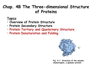
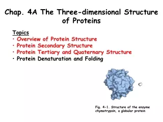
Chapter 6 The Three-Dimensional Structure of Proteins. Homework: 1, 9,12 ,. Proteins: Higher Orders of Structure . The structural variety of human proteins reflects the sophistication and diversity of their biologic role

E N D
Chapter 6 The Three-Dimensional Structure of Proteins Homework: 1, 9,12,
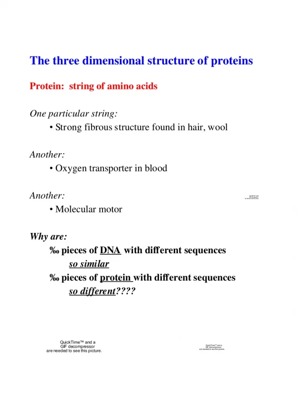
Proteins: Higher Orders of Structure • The structural variety of human proteins reflects the sophistication and diversity of their biologic role • The maturity of a newly synthesized polypeptide into a biologically active protein requires that it be folded into a specific 3-D arrangement or conformation
Conformation vs. Configuration • Configuration deals with the arrangement of specific bonds about individual atoms • Conformation refers to the spatial relationship of every atom of a molecule • Proteins were initially characterized by their gross characteristics
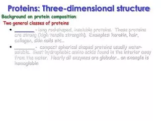
Initial Characterizations • Soluble proteins • Globular proteins • Fibrous proteins • Lipoproteins • Glycoproteins • Metalloproteins
Currently classification and Folding • Now, proteins are classified based on similarity or Homology, of residue sequence and structure • Typical proteins could have >1050 possible conformations • But since they fold as they form, it is easier to achieve biologically active conformation
Orders of protein structure • Primary Structure, 1o • Secondary Structure, 2o • Tertiary Structure, 3o • Quaternary Structure, 4o
Secondary Structure • The number of possible secondary structures is restricted • The is only free rotation about 2 of the 3 bonds in the backbone • Phi angle • Psi angle
Alpha Helix • Figure 6.3a, page 164 • Both Phi and Psi angles are defined as well as distance it rises per turn • Complete turn averages 3.6 residues • R groups face outwards • Mostly only righthandeda-Helix seen in nature due to mostly L-amino acids used!
Stabilizing the helix • H-bonds • van der Waals in core • Proline is only found in 1st turn. Why? • When present elsewhere, disrupts helix and forms a bend • Most hydrophobic R groups are on one side and hydrophilic on the other. Effect?
The Beta Sheet • As opposed to the helix, the amino acids are extended to form a zigzag or pleated pattern • The R groups alternate up/down for a joining residues • Stabilized by H-bonds, but without proximity • Figure 6.3b, page 165
Types of Beta Sheets • Parallel- adjacent segments proceed in same directions as far as amino to carboxyl • Antiparallel- Adjacent chains proceed in opposite directions, as in 6.3b. • Most have a right hand twist, they are not flat • Depictions, see figure 6.16, page 179
Loops and Bends • Approximately half the residues in a protein are in either a helix or beta sheet. • The other half are in less ordered conformations such as Loops, Bends, etc. • Turns and Bends are short segments that connect secondary structures
Beta Turn • Beta turns involve four Amino Acid residues with the 1st residue H-bonded to the 4th residue resulting in a tight, 180 degree turn • Fig 6.18 page 181 • Proline and Glycine are often present in beta turns. Why?
Loops • Loops are regions that contain more AA residues than minimally required to connect adjacent secondary structures • Although they have no set conformation, they do serve key biological roles • There are some conformations that are secondary structures interacting, but not enough to be termed a tertiary
Super Secondary Structures • Helix-Loop-Helix • Provide the oligonucleotide-binding portion of DNA-binding proteins such as repressors and transcription factors
Back to Loops • Loops are mainly found on the exterior portion of a protein. • Exposed to solvent • Readily accessible sites for recognition and binding • While the structure may be irregular, they still exist in specific conformations • Conformations are held together by H-bonds, salt bridges, and hydrophobic interactions
Tertiary and Quaternary Structure • 3o refers to the entire 3D conformation of the protein • Domain-a section of protein structure sufficient to perform a particular chemical or physical task such as binding of a substrate or ligand • Proteins may have one or more domains. • Fig 6.27 page 192
Tertiary and Quaternary Structure • In some cases, proteins are assembled from more than one polypeptide or protomer • Quaternary structure defines both the composition and spatial relations between these multiple chains.
Designations • Monomeric proteins consists of one chain • Dimeric proteins consists of two chains • For Dimeric and above, there are different kinds: • Homodimers-contain two copies of same chain • Heterodimers- contain two different chains
Designations • Greek letters are used to identify the different chains and subscripts indicate the number of each unit.
Stability of 3o and 4o structures • Primarily stabilized by non-covalent forces • Hydrophobic interactions- • H-Bonds • Salt-bridges • Etc • Think Velcro!!!
Determining 3D Structure • Two main techniques: • X-Ray crystallography • NMR Spectroscopy
X-Ray Chrystallography • Key is to get a good crystal!!! • Then bombard the crystal with x-rays and acquire a diffraction pattern • The diffraction pattern, along with the primary structure can be used to deduce the 3D structure.
X-Ray Chrystallography • Computers are getting better and better at interpreting X-ray diffraction patterns • Major stumbling block remains the requirement of inducing highly purified samples of the protein to crystallize • Several lines of evidence, including the ability of some crystallized enzymes to catalyze chemical reactions, indicate that the majority of the structures determined by X-ray crystallography represent the structures of proteins in free solution.
NMR Spectroscopy • Many common nuclei can be “seen” by NMR • Chemical shifts depend on the nuclei, what functional group it is in, and neighboring NMR nuclei
2-D NMR • Shows which nuclei are near each other.
Protein Folding • We have said the number of possible conformations of even small peptides is very large • Thermodynamics dictate the formation of the conformation • The desired conformation is usually energetically favored
Protein Folding • Even with this energetically favored situation, it would take billions of years for a protein to explore all possible conformations to find the favored state • So folding has to take place in some sort of guided environment • First, as protein leaves the ribosome, short segments fold into secondary structures which are directed by the primary sequence
Protein Folding • Second, the aqueous environment of the cell drives all hydrophobic side groups to the middle of the protein creating a molten globule • Regions of secondary structure rearrange to form the mature conformation. • While this process is ordered, it is not rigid
Definitions • In vivo- in the cell • In vitro- in test tube • Denatured- unfolding of a protein • Aggregates- disordered complexes of unfolded or partially unfolded polypeptides held together by hydrophobic interactions
Chaperones • Chaperone proteins help fold over half the proteins in mammals • They work by covering up hydrophobic groups, shielding them from the aqueous environment, thus preventing aggregation • Chaperons can also rescue misfolded proteins.
Higher Orders • Disulfide bonds help stabilize tertiary and quaternary structures even though they are non-specific. • The protein disulfide isomerase is present to continually break and reform disulfide bonds.
X-Proline Peptide Bonds • All X-proline peptide bonds-where X represents any residue-are synthesized in the trans configuration. • However, of the X-Proline bonds of mature proteins, approximately 6% are cis. • The cis configuration is particularly common in Beta-turns. • Isomerization from trans to cis is catalyzed by the enzyme proline-cis,trans-isomerase.
Alzheimer’s Disease • Refolding or misfolding of protein endogenous to human brain tissue, b-amyloid, is a prominent feature of Alzheimer’s Disease. • Levels of b-amyloid become elevated, and this protein undergoes a conformational transformation from a soluble helix-rich state to a state rich in beta sheets and prone to self-aggregaton.
Mass Spectrometry • Separates molecules based on MW • Peptides are vaporized • Applied charge propels the molecules down a bent tube • Molecules interact with magnetic field in tube • Where the molecules hit the side of the tube, which is the detector, gives their mass
Mass Spectrometry • Since all A.A. except Leu and Isoleu have different masses, all AA present can be determined • Post translational modifications can also be determined with this method
Advances • The methods of vaporization vary and recent advances have allowed for larger peptides to be used. • Book discusses “Time of Flight” MS.
Make sure you read!! • Make sure you read Ch. 6. Remember, you are responsible for material not covered in lecture. • Example: • Fibrous Proteins: Structural materials of Cells and tissue. Be able to compare and contrast Karatins, Fibron, collagen, and elastin.