Download
1 / 16
480 likes | 2.9k Views
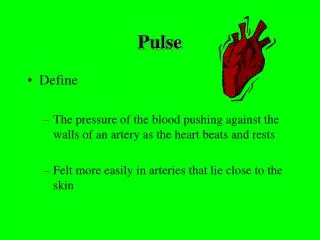
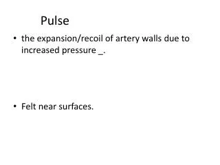
Pulse. LEQ: How does pulse differences aid in the diagnosis of a patient?. What is a pulse?. Pulse is defined as “the pressure of the blood pushing against the wall of an artery as the heart beats and rests.”

E N D
Pulse LEQ: How does pulse differences aid in the diagnosis of a patient?
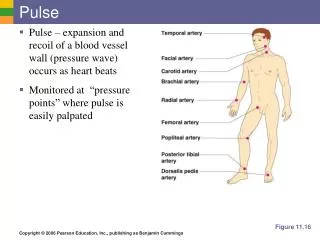
What is a pulse? • Pulse is defined as “the pressure of the blood pushing against the wall of an artery as the heart beats and rests.” • It is a throbbing of the arteries that is caused by the contractions of the heart. The pulse is more easily felt in arteries that le fairly close to the skin and can be pressed against a bone by the fingers.
Major sites pulses are felt • Temporal: at the side of the forehead • Carotid: at the neck • Brachial: inner aspect of the forearm at the antecubital space (crease of the elbow) • Radial: at the inner aspect of the wrist, above the thumb • Femoral: at the inner aspect of the upper thigh. • Popliteal: behind the knee • Dorsalispedis: at the top of the foot arch
APICAL PULSE • Apical pulse is a pulse count taken with a stethoscope at the apex of the heart. • The actual heartbeat is heard and counted.
What is a stethoscope? • A stethoscope is an instrument used to listen to internal body sounds. • The stethoscope amplifies the sounds so they are easier to hear.
Parts of a stethoscope • Use the smaller bell for a more focused exploration of sound in a smaller area. The smaller bell also gives more detail by reducing the area covered and shutting out nearby sounds that a larger bell would pick up. Use the smaller bell to distinguish between subtle sounds in specific locations like a cardiologist or other medical staff. Use the other side of the bell (with the diaphragm) to listen to a less focused area inside of the chest cavity.
Normal pulse ranges • Adult: 60- 100 • Infants: 100 to 160 • Infants pulse rate is higher because infant's body is growing rapidly and their body metabolism is high which needs more oxygen and to get ride of CO2 from the body quickly. This is achieved by increase in heart rate.
Abnormal pulse ranges • Tachycardia: pulse over 100 (except in children) • Bradycardia: pulse rate under 60 per minute. • Arryhytmia: irregular or abnormal rhythm, usually caused by a defect in the electrical conduction pattern of the heart.
Pulse deficit • Condition that occurs with some heart conditions. • In some cases, the heart is weak and does not pump enough blood to produce a pulse. In other cases, the heart beats too fast (tachycardia), and there is not enough time for the heart to fill with blood; therefore, the heart does not produce a pulse during each beat. • In these cases, the apical pulse rate is higher than the pulse rate at other pulse sites on the body.
How to check for a pulse deficit • For the most accurate pulse deficit determination, one person should check the apical pulse while a second person checks another pulse site. • If there is only one person, they should first check the apical pulse and then immediately check the radial pulse. • Then, subtract the rate of the radial pulse from the rate of the apical pulse. The difference is the pulse deficit. • Example: apical pulse is 120, the radial pulse is 80, the pulse deficit is 40.
http://www.easyauscultation.com/heart-sounds.aspx?gclid=CMLWh_bpwa0CFQtZ7AodBkyJBwhttp://www.easyauscultation.com/heart-sounds.aspx?gclid=CMLWh_bpwa0CFQtZ7AodBkyJBw • http://depts.washington.edu/physdx/heart/demo.html
Clinical: • Take 2 students pulse rates at rest and after activity. • Take their temporal, carotid, brachial, radial. • Count for a full minute. • Then get a stethoscope and listen for the apical pulse.