Download
1 / 33
330 likes | 343 Views
Malignant ovarian tumours. DR.FATIN. It is more common in the wealthy nations of the world. While the incidence is similar to that of endometrium &of cervix, more women die from ovarian cancer than from carcinoma of the cervix &body of the uterus combined because most of it discovered late.

E N D
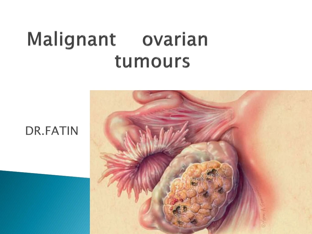
Malignant ovarian tumours DR.FATIN
It is more common in the wealthy nations of the world. While the incidence is similar to that of endometrium &of cervix, more women die from ovarian cancer than from carcinoma of the cervix &body of the uterus combined because most of it discovered late. Most of ovarian tumors are of epithelial origin.
Rare before 35years&incidence increase with age to a peak in the 50-70years old age group. Only 3% of ovarian cancers are seen in women younger than 35years &most of these are non epithelial cancer such as germ cell tumours.
Aetiology: Incessant ovulation theory: More in nulliparity Early menarche & late menopause(high estimated no. of years of ovulation) Oral contraceptive use reduce the risk 4 folds. Subfertility treatment: Subfertility, especially when it is unexplained is associated with both ovarian & longed attempts of ovulation induction.
Genetic factors: Familial ovarian cancer Forms 5-10% of women with ovarian cancer. Might associate with epithelial ovarian cancer (usually serous adenocarcinoma). Cases usually diagnosed before 50 years of ages. Defective genes include BRCA1 (81%) &BRCA2 (14%). The risk of ovarian cancer (40%) in these families which is less than the risk of breast cancer (80%). Genetic testing cannot guarantee to detect all defective genes. Annual ovarian u/s with color-flow Doppler studies & serum CA 125 estimation every 6-12 months are recommended.
Simplified histological classification of ovarian malignancy: 1.epithelial Serous carcinoma. Mucinous carcinoma. Endometrioid carcinoma. Clear cell carcinoma(mesonephroid) 2. Sex cord stromal tumours Granulosa cell tumours Androblastoma:Sertoli –Leyding cell tumours Gynandroblastoma.
3-Germ cell tumours Dysgerminoma Endodermal sinus tumours Choriocarcinoma Teratoma Mixed tumours 4-Metastatic tumours
Stages of Ovarian Cancer Stage I: This is the earliest form of ovarian cancer. Cancer is found in one or both ovaries. Stage I ovarian cancer is divided into three stages. Stage IA: Cancer is present in one ovary. Stage IB: Cancer is present in both ovaries. Stage IC: Cancer is present in one or both ovaries and cancer is found on the outside surface of one or both ovaries, or the outer covering of the tumor has ruptured, or cancer cells are found in the fluid or tissue linings of the abdomen.
Stage II:In stage II ovarian cancer, cancer is present in one or both ovaries, and has spread to other parts of the pelvic region. There are three stages in stage II ovarian cancer. Stage IIA: Cancer has spread to the uterus and/or fallopian tubes. Stage IIB: Cancer is in one or both ovaries and has spread to other organs in the pelvic region such as the bladder, rectum, or sigmoid colon. Stage IIC: Cancer is found in one or both ovaries and has spread to the uterus, fallopian tubes, bladder, sigmoid colon, or rectum. Cancer may also be present in tissue and fluid samples of the lining of the abdominal cavity.
Stage III: In stage III ovarian cancer, cancer is found in one or both ovaries and has spread to the abdomen. Stage III ovarian cancer is divided into three different stages. Stage IIIA: Cancer is found in one or both ovaries and has spread to a small part of the abdomen. Stage IIIB: Cancer is present in one both ovaries and has spread to the peritoneum in an amount less than 2 centimeters. Stage IIIC: Cancer is found in one or both ovaries, and has spread to the pertinoneum more than 2 centimeters and/or has spread to the lymph nodes.
Stage IV: Stage IV ovarian cancer is the most advanced stage of the disease. In this stage, cancer is found in one or both ovaries and has spread to parts of the body beyond the abdomen, like the lungs and liver.
Metastatic spread 2/3 of patient with ovarian cancer present with disease that has spread beyond the pelvis. 1.direct spread 2.peritoneal fluid (large&smallbowel,parietal peritoneal surface,liver) 3.lymphatic spread(pelvic¶-aorticlymphnodes,cervical nodes) 4.haematogenous spread :usually occurs late(liver,lung,bone,brain).
Ovarian Cancer Symptoms Most common symptoms: Bloating, abdominal pressure and/or discomfort Lower abdominal and/or pelvic pain Frequent and/or urgent urination (in absence of an infection) Feeling full quickly and/or difficulty eating Additional possible symptoms: Vague, but persistent gastrointestinal upsets (gas, nausea, indigestion) Change in bowel habits (constipation, diarrhea) Abnormal bleeding, pain during intercourse Unusual fatigue, shortness of breath Unexpected weight loss or gain
Ovarian cancer is difficult to detect in its earlier stages. Symptoms are often associated with the location of the tumor and its impact on the surrounding organs. They tend to be non-specific and can mimic other conditions.
Management of women with a family history of ovarian cancer: this depends on the women age,reproductive plans and individual risk. Ideally women with a strong family history should be referred to clinical genetics for assessment of the family tree. If the pedigree suggests a hereditary cancer ,genetic testing for BRCA1 and BRCA2 may be offered. At present .screening is offered to women aged 35 and over. This is usually yearly TVU and CA125 however this strategy is not very sensitive or specific. Prospective studies are being carried out looking at 4-6 mn measurement of CA125. Prophylactic bilateral salpingooopherectomy has a role in patients who are found to be carrying a gene mutation and have complete their family.
Examination and investigations of patient with epithelial ovarian tumour: Proper pelvic and abdominal examination may reveal a fixed hard mass arising from the pelvis in combination with the presence of ascites , a diagnosis of ov. CA is highly likely. Ultrasoundis the most useful non invasive test of a suspected malignancy. US characterizes the morphology of the cyst,presence of bilateral tumours,ascites and omemtal deposit.
blood test(CBC,LFT,RFT) CXR is important to assess pleural fluid and groin should be examined for enlarged nodes. Sometimes:bariumenema,colonoscopy,IVP ,CT scan of the abdomin and MRI if bowelsymptoms are present or there is a possibility of a primary colorectal tumour.
CA125 is the most common and is elevated in over 80% of EOC.CA125 blood test is used to measure the level of cancer antigen 125, a substance in the blood that can increase when ovarian cancer is present. However, because the CA125 level can be elevated by other conditions, and is not always increased in ovarian cancer patients, it is not an effective screening tool. Most doctors use the CA125 after the diagnosis of ovarian cancer has been surgically confirmed, as it can be an indicator of persistent or recurrent disease. Paracenthesis and pleural fluid aspiration may be required.
Surgery: Provided the patient is fit for anesthesia ,surgery remain necessary for diagnosis , staging and treatment of EOC. radical surgery is TAH & SALPANGIOOOPHRECTOMY + INFRACOLIC OMENTECTOMY. Fertility sparing surgery may also be performed in young patients with early stage EOC. In these cases unilateral salpingooopherectomy ,omentactomy,peritoneal biopsies and pelvic /paraaortic node dissection can be performed with endomaterial sampling to exclude tumour. Second look surgery is a planned laparotomy at the end of chemotherapy. The main function is to assess and resect any residual disease.
Chemotheropy can be given as primary treatment ,as an adjunct following surgery or for relapse of disease. It can be used to prolong clinical remission and survival or for palliation. Carboplatin or cisplatin are platinum compounds are the most effective chemotherapeutic agent in OV. CA. Carboplatin is less renal toxic and causes less N and V than platinum so it is most commonly used. The dose is calculated according to GFR.
Paclitaxel works by causing microtubar damage to the cell. This prevents replication and cell division. Pre-emptive steroid are given due to high sensitivty reaction. Side effects of peripheral neuropathy, neutopenia and myalgia are common and dose dependant. It also causes loss of total body hair irrespective of dose. After chemotherapy ,the patient may have a further CT scan to assess the response. The follow up of patient is usually by clinical examination and CA125level assessment.
Prognostic factors in OV. CA: - Stage of disease. -Volume of residual disease post surgery. -histological type and grade of tumour. -age at presentation. 5 years survival for stage 1 is 70-90%. 5 years survival for stage 11is 80%. 5 years survival for stage 111 is 30%. And for 1V is 10-20%.
Non epithelail tumours: Constitute about 10% of all ovarian tumours. Sex cord stromal tumour: 1-Granulose and theca cell tumours: Which is the most common sex cord tumour. They produce steroid hormones in particular oestrogen which can cause postmenapausal bleeding in older women and sexual precocity in prepubertal girl. The surgical treatment is the same as for epithelial tumours. Unilateral oopherectomy is indicated in young women with stage 1a disease. 2-sertoli –leydig cell tumours: Half of these rare neoplasia produce male hormones which can cause virilization. The treatment the same of granulosa cell tumours.
Germ cell tumours: 1-dysgerminoma: Account for 2-5% of all primary malignant OV. Tumours. All occur in young women less than 30 years old. They spread mainly by lymphatic. All cases need CXR and CT . serum AFP and BHCG must be assayed to exclude the presence of choriocarcinoma ,endodermal sinus tumour or teratoma. • 2-yolk sac tumors: Also called endodermal sinus tumours making up 10-15% over all and reaching higher proportion in children. It may present as acute abdomine due to rupture of tumour following necrosis or hge. Often secrete AFP.
3-teratoma: Mature teratoma are benign ,the most common being the cystic teratoma or dermoid cyst found at all ages but particularly in the third and fourth decades. The treatment of all germ cell tumours mainly conservative surgery because the patients are usually young. Combination chemotherapy is highly effective when required.
Thank u……. Thank u……. Thank you……… Thank u…….