Download
1 / 54
560 likes | 811 Views
Myelodysplastic and Myelodysplastic / Myeloproliferative Neoplasms.

E N D
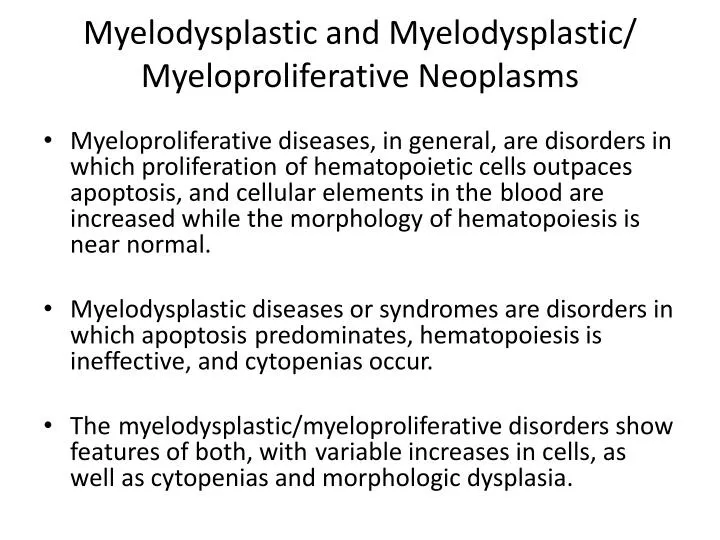
Myelodysplastic and Myelodysplastic/MyeloproliferativeNeoplasms • Myeloproliferative diseases, in general, are disorders in which proliferationof hematopoietic cells outpaces apoptosis, and cellular elements in theblood are increased while the morphology of hematopoiesis is near normal. • Myelodysplastic diseases or syndromes are disorders in which apoptosispredominates, hematopoiesis is ineffective, and cytopenias occur. • Themyelodysplastic/myeloproliferativedisorders show features of both, withvariable increases in cells, as well as cytopenias and morphologic dysplasia.
Types of Abnormal Cellular Maturation • Dyserythropoiesis : Nuclearfragmentation or karyorrhexis, multinuclearity, nuclear budding or bridging,basophilicstippling, and ring sideroblasts. • Erythrocyticabnormalities in the blood film include presence of oval macrocytes, anisochromia,basophilic stippling, dacryocytes, and reticulocytopenia.
Dysgranulopoiesis: hypogranulation,nuclearhyposegmentation with increased chromatin condensation,occasionally abnormal large azurophilicgranules. Dysmegakaryocytopoiesis: large megakaryocytes with unsegmentednuclei, micromegakaryocytes, and megakaryocytes with two ormore small, unconnected nuclei .Megakaryocytesmay bedecreased in number. In the blood film, giant hypogranular platelets arefrequent, and micromegakaryocytes are seen rarely.
A dysplastic megakaryocyte showing unconnected nuclear lobes
Myelodysplastic/MyeloproliferativeNeoplasms • Chronic MyelomonocyticLeukemia • Atypical Chronic Myeloid Leukemia,BCR-ABL1 Negative • Juvenile MyelomonocyticLeukemia • Myelodysplastic/MyeloproliferativeDisease,Unclassifiable
Chronic Myelomonocytic Leukemia • CMML is a clonal stem cell disorder in which the predominant feature ispersistent monocytosis (>1 × 10⁹/L for longer than 3 months) for whichother causes have been excluded. • Blast percentages typically are <5% in the blood and <10% in themarrow, and such cases are referred to as CMML-1. The subcategoryCMML-2 refers to cases with 5%–19% blasts in the blood, with 10%–19%in the marrow, or with the presence of Auer rods.
CMML • درCMML تقریبا همیشه بیشتراز 10% مونوسیت وجود دارد اما در CML به ندرت از 2-3% تجاوز می کند. • درCMML کروموزوم فیلادلفیا وجود ندارد و دیس پلازی یک یاچند رده میلوئیدی وجود دارد و کمتر از 20درصد سلولهای BM را بلاست و پرومونوسیتها تشکیل می دهد. • هرگاه تعداد ائوزینوفیلها از 1500/μlبیشتر گردد WHO زیرگروه CMML همراه با ائوزینوفیلی پیشنهاد می کند. • از نظر ایمونوفنوتیپ:CD13,CD33مثبت وCD14,CD64,CD68به صورت متغییر مثبت است. • ناهنجاریهای سیتوژنتیک در20-40% موارد +8,-7و ناهنجاریهای 12P می باشد، برخی از بیماران t(5,12) یا t(5,17) همراه با افزایش ائوزینوفیل وپاسخ درمانی مناسب به بازدارنده های تیروزین کینازی دارند. • میزان متوسط بقا در بیماران 20-30 ماه می باشد و 15-30 درصد پیشرفت به سمت AML دارند.
Atypical Chronic Myeloid Leukemia,BCR-ABL1Negative Criteria for diagnosis of aCML: • Leukocytosis(WBC ≤13 × 109/L) due to increased neutrophilsandprecursors with prominent dysgranulopoiesis • Prominent dysgranulopoiesis • No Ph′ chromosome or BCR/ABL-1 fusion gene • No rearrangement of PDGFRA or PDGFRB • Neutrophil precursors (promyelocytes, myelocytes, metamyelocytes)≥10% of WBCs • Minimal absolute basophilia; basophils usually <2% of leukocytes • No or minimal absolute monocytosis; monocytes <10% of WBCs • Hypercellular marrow with granulocytic proliferation and granulocyticdysplasia, with or without dysplasia of erythroid and megakaryocyticlineages • Less than 20% blasts in blood or marrow
Atypical CML. Top panels: Peripheral blood smear from a patient with atypical CML showed a leukocytosis with anemia and marked thrombocytopenia. Granulocytes are left-shifted, with dysplastic hypolobulated nuclei and a dysplastic nRBC is present (arrow). • Bottom panels: Biopsy at low- and high-magnification (left and right, respectively) showing atypical granulocytic hyperplasia without sheets of blasts.
Juvenile Myelomonocytic Leukemia • Juvenile myelomonocytic leukemia (JMML) is a clonal disorder of predominantlygranulocytic and monocytic lineages, with dysplasia in theseand frequently other lineages, occurring in children or young adolescents • The occurrence is 1.3 casesper million children younger than 14 years, and most affected children areyounger than 3 years of age, with a 2 : 1 male predominance. • JMML isfrequent in children withneurofibromatosis type 1.
In all, 25% of patients show monosomy 7, 35% exhibit mutations ofPTPN11 (encoding SHP2), and 20% each have mutations in NRAS,KRAS2, and NF1. • Patientswith normal karyotype often have markedly increased hemoglobin F. • Blood shows leukocytosis, thrombocytopenia, and often anemia. • The marrow is hyperplastic with granulocytichyperplasia, variable erythroidcellularity, and often decreasedmegakaryocytes.
Criteria for diagnosis of JMML • Peripheral blood monocytosis >1 × 10⁹/L • Absence of Ph′ chromosome or BCR/ABL-1 • Blasts and promonocytes less than 20% of blood and marrow • Plus two of the following: • Hemoglobin F increased for age • Immature granulocytes in the blood • WBCs >10 × 109/L • Clonalchromosomal abnormality (may be monosomy 7) • GM-CSF hypersensitivity of myeloid progenitors in vitro
This peripheral blood smear from a child with JMML shows dysplastic monocytosis, left-shifted leukocytosis with a small blast, and thrombocytopenia.
Myelodysplastic/Myeloproliferative Disease,Unclassifiable • This category of the WHO classification is used for those cases with featuresof myelodysplastic disease, but with the addition of prominentmyeloproliferative features. • Refractory Anemia with Ring SideroblastsAssociatedWith Marked Thrombocytosis
Refractory Anemia with RingSideroblasts AssociatedWith MarkedThrombocytosis • This disorder is included in the WHOclassification under the category ofMDS/MPD, unclassified. • It is characterized by features ofmyelodysplasticneoplasm andrefractory anemia with ring sideroblasts. • Peripheralthrombocytosis≥450 × 106/L, and increased, large atypical BM megakaryocytes. • A majority of cases (60%) carry a JAK2 V617F mutationidentical to that in MPN; occasional cases show a MPL W515K/Lmutation
Myelodysplastic Syndromes • اختلالات کلونال خونسازی هستند. • نارسایی BM علارغم سلولاریته بالای آن وجود دارد. • از نظر شیوع در مطالعات اروپایی3-4 نفر در100000تا اما با افزایش سن افزایش می یابد بطوریکه 30 نفر در 100000در سن بالای 80 سال میرسد نتایج در ایتالیا نشان داده که 60درصد مریضان سن بالای 70سال دارند. • 20 درصد بیماران با CBC اتفاقی شناسایی می شوند.اما اغلب با تصویری از نارسایی BM شناسایی می گردند. • شایعترین ناهنجاری کروموزومی در کروموزوم 5(FMS,FOS,RAS), 7(ERB-D), 8(MYC) که همگی Proto-oncogeneرا حمل می کنند، می باشد.موتاسیون RAS در 50% وموتاسیون FMS در 16%از MDSها گزارش شده است.
The Who classification of MDS • Refractory cytopeniawith unilineage dysplasia (anemia (RA),thrombocytopenia, or neutropenia) • Refractory anemia with ringed sideroblasts • Refractory cytopeniawith multilineage dysplasia • Refractory anemia with excess blasts (RAEB) • 5q− syndrome • Myelodysplastic Syndrome, Unclassified • Childhood MyelodysplasticSyndrome; Refractory Cytopenia of Childhood
Refractory Cytopenia with Unilineage Dysplasia • dysplasia affecting >10% of one myeloid cell lineage, and cytopenia of the affected cell line, with <10% dysplasia of other lineages. • Del (20q), +8, abnormalities ofchromosome5 or 7 • Refractory Anemia • Refractory Neutropenia • Refractory Thrombocytopenia
RA: • 5-10%از MDSها را تشکیل میدهد. • دیسپلازی مشخص وبارز مربوط به رده اریتروئیدی است. • آنمی، رتیکلوسیتوپنی، اریتروسیتهای غیرطبیعی، دایمورفیک،آنیزوپویکیلوسیتوز خفیف تا مشخصی وجود دارد. • درBM هایپرسلولاریتی معمول می باشد اما گاهی نرمو یا حتی هیپوسلولار می باشد.دیسپلازی اریتروئیدی از خفیف تا متوسط متغیر است. • میزان بلاست در PB کمتر از 1% و در BM کمتر از 5%ومیزان سیدروبلاست حلقویBM کمتراز15% اریتروبلاستها می باشد. • میزان بقا 66ماه و transformation به AML حدود 6 درصد می باشد.
Refractory Neutropenia • The absolute neutrophil count is <1.8 × 10⁹/L. • Toxic/secondary neutropeniais excluded. • Dysplasia usually consistsof nuclear hypolobation and hypogranulation. • Refractory Thrombocytopenia • Platelets are <100 × 109/L • More than 10% of at least 30 megakaryocytesevaluated in smears and sections show dysplastic features of hypolobation,binucleationor multinucleation, and/or micromegakaryocytes.
Refractory Anemia with Ring Sideroblasts • Similar to RA with >15% ringedsideroblastsin BM • Survival is similar to RA, with a lowerprogression to AL (≤2%).
Refractory CytopeniawithMultilineageDysplasia • PB with cytopenias of ≥2 cell lines,<1% blasts, • <1 × 10⁹/L monocytes. • BM with dysplasia of ≥10% ofprecursors of ≥2 cell lines, <5%blasts • Ring sideroblasts may be present;when numerous (>15%), the case is still classified as RCMD and is notcategorized separately, as in prior classifications. • Survival is ≈33months, and AL conversion is 11%
RA and RARS, which infrquently show cytogenetic abnormalities(<10%) • RCMD and RCMD with ring sideroblasts (RCMD-RS)show cytogenetic abnormalities in up to 50%. These include trisomy 8, monosomy 7, del(7q),monosomy 5, del(5q), del(20q)
Refractory Anemia with Excess Blasts • RAEB-1: PB with <5% blasts,<1 × 10⁹/L monocytes; BM withhypercellularity, dyspoiesis, 5%–9%blasts without Auer rods; Survival is usually less than 2 years(18month)in RAEB-1 • with progressive marrow failure andcytopeniasor progressionto AL in 25% • Clusters of 5–8 blasts and promyelocytes in themarrow interstitium are frequently seen on sections and have been referredto as abnormal localization of immature precursors(ALIP).
Cases of RAEB with concurrent myelofibrosis are referred to as RAEBwith fibrosis (RAEB-F). Megakaryocytes are increased and dysplastic,and reticulin fibrosis is significant, often resulting in a dry tap.
RAEB-2: • PB with 5%–9% blasts, or10%–19% BM blasts, or Auer rods • Survival is usually less than 2 years(10 month)in RAEB-2 • progressive marrow failure and cytopenias or progressionto AL in 33% for RAEB-2. • Cytogenetic abnormalities include +8, -5, del(5q), -7, del(7q), and del(20q)
Myelodysplastic Syndrome with Isolated del(5q) • PB with thrombocytosis, <5% blasts; • BM with increased, hypolobulatedmegakaryocytes, <5% blasts • isolated deletion of chromosome region 5q. • Some cases also exhibit a JAK2 V617F mutation.
5q-syndrome. blood smear showing macrocytic anemia and thrombocythemia. Macrocytic RBCs with cellular diameters that exceed the nucleus of a small mature lymphocyte are seen.
5q-syndrome. Aspirate smear show increased numbers of the characteristic unilobularmegakaryocytes.