Download
1 / 48
980 likes | 3.99k Views
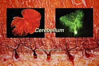
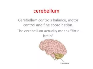
Cerebellum (Latin for Little Brain ). Function The cerebellum regulates the following 5 functions: Muscle tone Coordination of goal directed and spontaneous movements Posture and balance Eye movements Motor learning

E N D
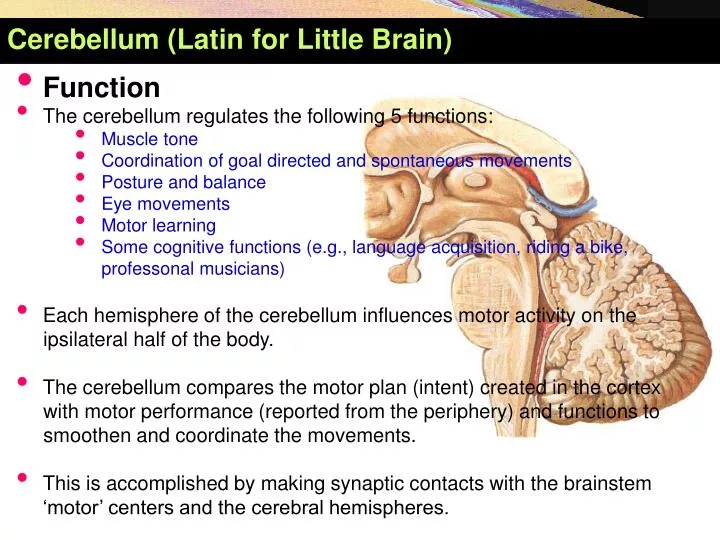
Cerebellum (Latin forLittleBrain) • Function • The cerebellum regulates the following 5 functions: • Muscle tone • Coordination of goal directed and spontaneous movements • Posture and balance • Eye movements • Motor learning • Some cognitive functions (e.g., language acquisition, riding a bike, professonal musicians) • Each hemisphere of the cerebellum influences motor activity on the • ipsilateralhalf of the body. • The cerebellum compares the motor plan (intent) created in the cortex • with motor performance (reported from the periphery) and functions to • smoothen and coordinate the movements. • This is accomplished by making synaptic contacts with the brainstem ‘motor’ centers and the cerebral hemispheres.
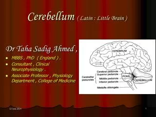
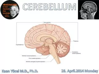
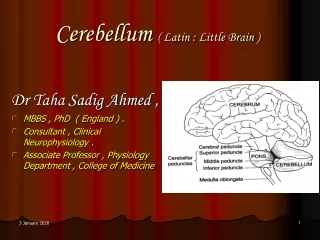
TheCerebellum • Its surface is highly convoluted, forming folds or folia, being oriented transversely. • Lies behind pons & M.O. , separated from them by the cavity of 4th ventricle. • It is connected to brain stem (medulla, pons& midbrain) by inferior, middle & superior cerebellar peduncles, respectively.
Cerebellum, SuperiorandInferiorSurfaces • The cerebellum consists of a midline vermisand2lateral hemispheres. • Anatomically,it is divided intoanterior, posterior & flocculo-nodular lobes. Vermis: (Latin for worm)
External Features of Cerebellum • It has anterior notch,which is wider and lodging the back of pons & medulla. It is separated from them by cavity of 4thventricle. • It has also posterior notch occupied by falx cerebelli, which separates the 2 cerebellar hemispheres.
External Features of Cerebellum • Superior surface: lies beneath tentorium cerebelli and has a raised superior vermis+ a large cerebellar hemisphere on each side + primary & horizontal fissures. • Primary fissure V-shaped,well defined fissure, lies on superior surface and separates the small anterior lobe from the larger middle lobe (or posterior lobe). • Horizontal fissure lies along the sides of cerebellum, extending from anterior notch to posterior notch, separates the superior from the inferior surfaces.
External Features of Cerebellum • Inferiorsurface:rounded oneach side and presents : • Adeep groove (vallecula) between 2 cerebellarhemispheres,which is occupied by the inferior vermisandtonsil. • Tonsil isasmall part of cerebellar hemisphere that lies lateral to inferior vermis. • Retrotonsillarfissureseperatestonsilfromthebiventerlobule. • Secondary (posterolateral) fissurelies on inferior surface and separates flocculo-nodular lobe from the remainder of cerebellum.
External Features of Cerebellum Uvula: (Latin for little grape) Tuber: (Latin for swelling)
Internal Features of Cerebellum Interposed Nucleus
CerebellarNuclei • Fastigial • Stance and gait, controls muscles only in the modes of sitting, standing, and walking • Globose + Emboliform • Segmental reflexes, speeds the initiation of movements triggered by somatosensory cues that guide the response, stops unwanted and promotes wanted oscillations, stabilizes holds • Dentate • Fine dexterity
Functional Subdivisions of Cerebellum • Schematic drawing of cerebellum showing the relationships between the anatomical & functional divisions of cerebellum. (Paleo-cerebellum) (Neo-cerebellum) (Archi-cerebellum)
Functional Subdivisions of Cerebellum • 1- Archi-cerebellum = posterior lobe (Vestibular part) : • It is formed of the flocculo-nodular lobe + associated fastigial nuclei, lying on inferiorsurface in front of postero-lateral fissure. • Embryologically, it is the oldest part of cerebellum. • It receives afferent fibers from vestibular apparatus of internal ear via vestibulocerebellar tracts. • It is concerned with equlibrium and eye movements
Functional Subdivisions of Cerebellum • Archicerebellum • It is concerned with equilibrium. • It represents flocculo-nodular lobe. • It has connections with vestibular & reticular nuclei of brain stem through the inferior cerebellar peduncle. • Afferent vestibular fibers. Pass from vestibular nuclei in pons & medulla to the cortex of ipsilateral flocculo-nodular lobe. • Efferent cortical (purkinje cell) fibers. Project to fastigial nucleus, which projects to vestibular nuclei & reticular formation. • It affects the L.M.system bilaterally via descending vestibulo-spinal & reticulo-spinal tracts. Connections of archicerebellum
Functional Subdivisions of Cerebellum • 2. Paleo-cerebellum= (spinal part) :Formed of: • Midline vermis + surrounding paravermis + globose & emboliform nuclei. • It receives afferentproprio-ceptive impulses from Ms.& tendons viaspino-cerebellar tracts (dorsal & ventral) mainly. • It sends efferents tored nucleus of midbrain. • It is concerned withmuscle tone.
Functional Subdivisions of Cerebellum • 2. Paleo-cerebellum • It is concerned with muscle tone & posture. • Afferents spinal fibers: consist of dorsal & ventral spino-cerebellar tractsfrom muscle, joint & cutaneous receptors to enter the cortex of ipsilateral vermis & para vermis via inferior & superior cerebellar peduncles. . • Efferents cortical fibers pass to globose & emboliform nuclei, then via sup. c. peduncle to contra-lateral red nucleus of midbrain to give rise descending rubro-spinal tract. Connections of Paleo-cerebellum.
Functional Subdivisions of Cerebellum • 3- Neo-cerebellum= (cerebral part): • It is the remaining largest part of cerebellum. • It includes the most of 2cerebellarhemispheres + dendate nuclei. • It receives afferent impulses from the cerebral cortex+pons via cerebro-ponto- cerebellar pathway. • It sends efferents to V.L.nucleus of thalamus. • It controls voluntary movements (muscle coordination).
Functional Subdivisions of Cerebellum • 3- Neo-cerebellum • It is concerned withmuscular coordination. • It receives afferents from cerebral cortex involved in planning of movement- to pontine nuclei,cross to opposite side via middle cerebellar peduncle to end in lateral parts of cerebellum(cerebro-ponto-cerebellar tract). • Neo-cerebellar efferents project to dendate nucleus,which in turn projects to contra-lateral red nucleus & ventral lateral nucleus of thalamus,then to motor cortex of frontal lobe, giving risedescending cortico-spinal & cortico-bulbar pathways. • Efferents of dentate nucleus form a major part ofsuperior C. peduncle. Connections of Neo-cerebellum.
Afferent and Efferents • Superior Cerebellar Peduncle (brachium conjunctivum) • Connects with midbrain • Middle Cerebellar Peduncle (brachium pontis) • Connects with pons • Inferior Cerebellar Peduncle (restiform body) • Connects with medulla
Superior Cerebellar Peduncle • Afferent: • Ventral spinocerebellar tract: Transmits proprioceptive and exteroceptive information from levels below the midthoracic cord. • , Tectocerebellar tract: Arises in the superior and inferior colliculi and carries auditory and visual information. • Trigeminocerebellar tract: Carries proprioceptive fibers from the mesencephalon and tactile information from the chief sensory nucleus of the trigeminal nerve. • The cerulocerebellar tract: Fibers from the nucleus ceruleus
Superior Cerebellar Peduncle Efferent: (most prominent) • Dentatorubral tract, output to the contralateral red nucleus • Dentatothalamic tract, output to the contralateral ventrolateral nucleus of the thalamus • Uncinate bundle of Russell, output to the vestibular nuclei and reticular formation
Middle Cerebellar Peduncle • Afferent fibers: (only) • Pontocerebellar (corticopontocerebellar) tract, arises in contralateral pontine gray matter • Transmits impulses from the cerebral cortex to the intermediate and lateral zones of the cerebellum.
Inferior Cerebellar Peduncle Afferents: • Dorsal spinocerebellar tract, originating in the dorsal nucleus of Clarke (T1–L2), carries proprioceptive and exteroceptive information mostly from the trunk and ipsilateral lower extremity. • Cuneocerebellar tract, originating in the external arcuate nucleus, which transmits proprioceptive information from the upper extremity and neck. • Olivocerebellar tract, which carries somatosensory information from the contralateral inferior olivary nuclei. • Vestibulocerebellar tract, transmits information from vestibular receptors on both sides of the body. • Reticulocerebellar tract, arises in the lateral reticular and paramedian nuclei of the medulla.
Inferior Cerebellar Peduncle • Arcuatocerebellar tract, arises from the arcuate nuclei of the medulla oblongata. • Trigeminocerebellar tract, arises from the spinal and main sensory nuclei of the trigeminal nerve Efferent: • Fastigiobulbar tract -(through Juxtarestifrom body), cerebellovistibular • Cerebelloreticular pathways
A schematic model of the motor system. The cerebellum influences movements via connections to both the brainstem and cerebral cortex
Vascular Supply • PICA • From intracranial vertebral artery, supplies the lateral medullary tegmentum, inferior cerebellar peduncle, the ipsilateral portion of the inferior vermis, and the inferior surface of the cerebellar hemisphere. • Medial branch supplies dorsolateral medulla and medial cerebellum, lateral branch supplies inferiopostalateral
Vascular Supply • AICA • Above the origin of the basilar artery, supplies the anterior petrosal surface of the cerebellar hemisphere, flocculus, lower portion of the middle cerebellar peduncle, and lateral pontomedullary tegmentum. • SCA • Distal segment of the basilar artery just below the terminal bifurcation into the paired PCAs, and supplies the upper surface of the cerebellar hemisphere, ipsilateral portion of the superior vermis, most of the dentate nucleus, upper portion of the middle cerebellar peduncle, superior cerebellar peduncle, and lateral pontine tegmentum.
CerebellarLesions • Are usually vascular, may be traumatic or tumor. • Manifestations of unilateral cerebellar lesions : • Ipsilateral incoordination of (U.L) arm = intention tremors : it is a terminal tremors at the end of movement as in touching nose or button the shirt. • Ipsilateral cerebellar ataxia affects (L.L.) leg, causing wide-based unsteady gait. • Manifestations of bilateral cerebellar lesions (caused by alcoholic intoxication, hypothyrodism, cerebellar degeneration & multiple sclerosis) : • Dysarthria: slowness & slurring of speech.
CerebellarLesions • Incoordinationof both arms.= intention tremors • Cerebellar ataxia: intermittent jerky movements or staggering , wide-based, unsteady gait. • Nystagmus :It is due to impairment coordination of eye movements /so, incoordination of eye movements occurs and eyes exhibit a to-and-fro motion. • Combination of nystagmus+ dysarthria+ intension tremors constitutes Charcot’s triad, which is highly diagnostic of the disease. • Loss of cerebellar function does not produce paralysis or the inability to initiate a movement. Rather, cerebellar disease produces disturbances in the coordination and fine control of movements and posture.