Download
1 / 1
10 likes | 182 Views
A RARE CAUSE OF RECTAL PAIN Irene Krokos MD, John R. Pierce, MD University of New Mexico School of Medicine. Extrapulmonary Tuberculosis. Anorectal Tuberculosis. Case Presentation

E N D
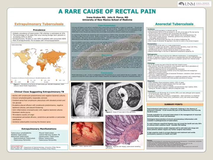
A RARE CAUSE OF RECTAL PAIN Irene KrokosMD, John R. Pierce, MD University of New Mexico School of Medicine Extrapulmonary Tuberculosis Anorectal Tuberculosis Case Presentation A 64 year-old Hispanic male without a significant past medical history presented with a complaint of “rectal pain.” The pain was worse with bowel movements and as a result the patient reported a fear of eating. The patient denied any associated nausea, vomiting, fevers, chills, melena or bright red blood per rectum. He did report a two week history of a dry nonproductive cough. Additionally he had a 50 pack-year smoking history and multiple sexual partners without consistent use of protection. On examination the patient was hypotensive with a blood pressure of 80/60 mmHg, tachycardic and had a decrease in weight from 60kg to 45kg in less than 6 months. Breath sounds were clear bilaterally and rectal examination revealed a 4 cm by 4 cm rectal ulceration. Stool exam was brown and guiac positive. Laboratory examination revealed a mild leukocytosis with normal differential and chest radiology showed diffuse patchy opacifications. Given the patient’s social history, weight loss and chest radiology findings there was concern for human immunodeficiency virus (HIV) infection and Pneumocystis jiroveci pneumonia. He was started on trimethoprim-sulfamethoxazole and admitted for further evaluation. The patient’s hypotension responded to fluids. He was evaluated by gastroenterology and infectious disease services. HIV testing was negative. A new working diagnosis of rectal malignancy with lymphangitic spread to the lungs was made. The patient underwent colonoscopy with biopsies. Sigmoid ulcerations were also noted. Rectal biopsies revealed caseating granulomas and 2+ acid fast bacilli (AFB). Induced sputum later showed 4+ AFB. The patient was placed in isolation and started on a four drug regimen for treatment of miliary tuberculosis (TB). Discussion National TB surveillance data reveals that almost one-fifth of TB cases in the United States are extrapulmonary. Gastrointestinal TB is a diagnostic challenge in the absence of a pulmonary infection. Only 2% of gastrointestinal TB cases present after 60 years of age. Most commonly the intestinal lesions are ulcerative. Symptoms include abdominal pain, diarrhea, weight loss, fever, melena and rectal bleeding. Rectal lesions usually present as anal fissures, fistulas or perirectal abscesses. It is essential to distinguish TB enteritis from inflammatory bowel disease such as Crohn’s disease as the initiation of immunosuppressive therapy in a patient with tuberculosis can lead to dissemination. Our patient presented with rectal involvement and likely had disseminated TB. Classic miliary TB is defined as millet like seeding of TB bacilli in the lung and is seen in 1-3% of all TB cases. It can mimic many diseases and in some cases up to 50% are diagnosed ante mortem. A high index of clinical suspicion is important as early diagnosis and treatment correlate with improved outcomes. Conclusions Rectal tuberculosis is rare. A case of undiagnosed rectal TB presenting as an acute perianal abscess is reported. Lack of suspicion for rectal TB in such a case can lead to delays in diagnosis and significant risks of exposure to healthcare personnel. • Incidence: • One-third world population is infected with TB • Extrapulmonary forms of TB are present in 10-15% of all cases of TB, but can be • found in 40 to 60% of patients with concomitant HIV infection • Gastrointestinal (GI) tract is the 6th most frequent site of extrapulmonary TB • (From most to least common: lymphatic, genitourinary, bone/joint, miliary, • meningeal, gastrointestinal) • Anorectal TB compromises less than 2% of cases of abdominal TB • Only 3 cases of anorectal TB reported in the last 22 years in the United Kingdom • Presentation: • Fourth decade of life with a 4:1 male predominance • Symptoms: weight loss (40-90%), abdominal pain (80-95%), fever (40-70%), • change in bowel habits (50%), anorexia and malaise • Hematochezia (due to mucosal trauma by stool) is common (88%) • Massive hemorrhage is rare due to obliterative endarteritis caused by TB • Pathogenesis: • TB bacilli can reach the GI tract by four different mechanisms: 1) hematogenous spread, 2) ingestion of bacilli from sputum or unpasteurized milk from infected bovine, 3) direct spread from adjacent organs and 4) lymphatic spread from infected lymph nodes • Pathology: • GI TB can involve any part of the GI tract from mouth to anus • Most common site of GI involvement is the ileocecal region due to abundance of • lymphoid tissue (M cells and Peyer’s patches) • Ulcers are superficial and do not penetrate the muscularis • They tend to be transversely oriented versus the longitudinal and serpiginous • appearance of Crohn’s ulcers • There are 4 morphological types of anorectal TB lesions: ulcerative (most common), • verrucous, lupoid and miliary • The ulcerative form typically presents as a superficial ulceration with a hemorrhagic • necrotic base that is covered with thick purulent secretions of mucous • Diagnosis: • Chest radiology shows pulmonary lesions <25% of cases • Colonoscopy requires multiple biopsies from the ulcer edge • Cultures are positive in 40% of biopsies • Acid-fast bacilli (AFB) staining is variable and polymerase chain reaction (PCR) • testing for TB DNA is helpful in difficult to diagnose cases • Treatment: • Conventional anti-TB therapy for 6 months (99% cure rate) although some expand • to 12-18 months (94% cure rate) • Surgery is indicated only if there is a complication. Most commonly it is intestinal • obstruction (15-60%), fistula (25%), perforation (15%) and rarely hemorrhage Prevalence • Globally prevalence of tuberculosis (TB) infection is estimated at 32% • The percentage of US cases that occur among foreign-born persons is • increasing (53% in 2003) • Extrapulmonary TB seen in over 50% of patients with concurrent AIDS • Risk of extrapulmonary TB increases with immunosuppression Figure 1 – Prevalence of all forms of tuberculosis per 100,000 inhabitants, 2005 (Source: World Health Organization) Clinical Clues Suggesting Extrapulmonary TB • Ascites with lymphocyte predominance and negative bacterial cultures • Chronic lymphadenopathy (especially cervical) • Cerebral spinal fluid lymphocytic pleocytosis with elevated protein and • low glucose • Exudative pleural effusion with lymphocyte predominance, negative • bacterial cultures, and pleural thickening • Joint inflammation (monoarticular) with negative bacterial cultures • Persistent sterile pyuria • TB-endemic country of origin • Unexplained pericardial effusion, constrictive pericarditis or pericardial • calcification • Vertebral osteomyelitis involving the thoracic spine SUMMARY POINTS Gastrointestinal tuberculosis is a diagnostic challenge in the absence of pulmonary infection. It can involve any part of the alimentary tract from the mouth to anus. Include evaluation for anorectal tuberculosis in the management of recurrent anorectal fistulas, ulcers and abscesses. Histological demonstration of chronic granulomatous inflammation with caseation is pathognomonic of tuberculosis. In most instances superficial biopsies may not reveal the bacilli and only 36% of cultures yield a positive result. Consider PCR testing. Anorectal tuberculosis usually responds well to anti-tubercular drugs and these patients seldom require any further surgical intervention. A high suspicion leads to prompt diagnosis and treatment and avoids unnecessary exposure to health care staff. Figure 3 - Chest CT scan later in the admission Figure 2 - Chest X-ray on initial presentation Extrapulmonary Manifestations TuberculousLymphadenitis Pleural Tuberculosis Skeletal Tuberculosis Central Nervous System TB Abdominal Tuberculosis Genitourinary Tuberculosis Gastrointestinal Tuberculosis Milliary Tuberculosis Tuberculous Peritonitis TuberculousPericarditis References: 1) Samarasekera, DN, Nnayakkara, PR. Rectal tuberculosis: a rare cause of recurrent rectal suppuration. Colorectal Disease. 2008: 846-848. 2) Kamani L, et al. Rectal tuberculosis: the great mimic. Endoscopy 2007: E277-E228. 3) Golden, MP, Vikram, HR. Extrapulmonary tuberculosis: an overview. American Family Physician 2005: 1761-1768. 4) Sharma, MP, Bhatia, V. Abdominal tuberculosis. Indian Journal of Medical Research 2004: 305-315. 5) Saenz, EV, et al. Colonic tuberculosis. Digestive Diseases and Sciences. 2002: 2045–2048. 6) Subnis, BM, et al. Primary tuberculosis of rectum mimicking malignancy: a case report. Bombay Hospital Journal. 2008: 283-285. Acknowledgements Michael Gilles, MD – Department of Gastroenterology, University of New Mexico Mark Hubbell, MD – Department of Pathology, University of New Mexico Figure 4 – Colon, sigmoid ulcer Figure 5 – Sigmoid ulcer biopsy, submucosal caseating granuloma