Download
1 / 1
10 likes | 134 Views
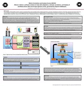
Optical Nanoparticle Trapping Sensor . Gaussian Beam Shape. Dispersion. Evanescent Field Decay. F GRAD. Darryl Benally †, Dr. Matt Kipper*‡, Dr. Randy Bartels†‡

E N D
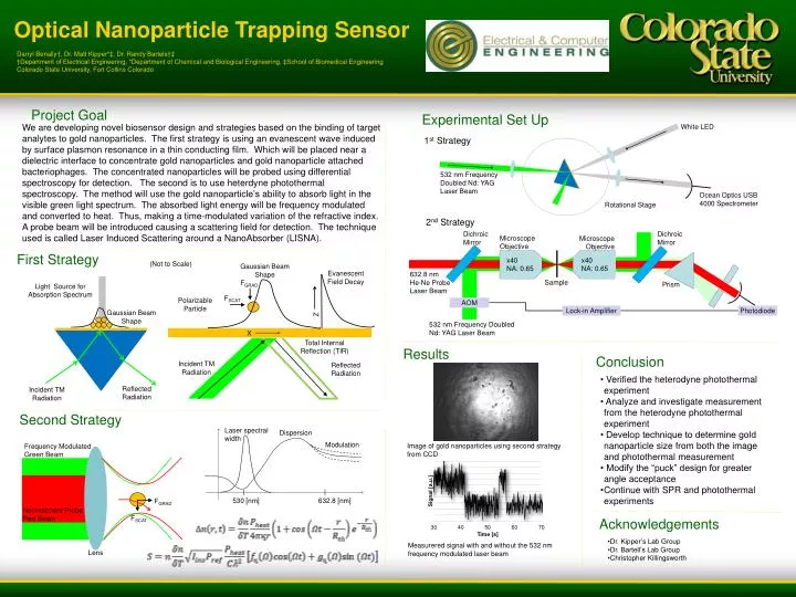
Optical Nanoparticle Trapping Sensor Gaussian Beam Shape Dispersion Evanescent Field Decay FGRAD Darryl Benally†, Dr. Matt Kipper*‡, Dr. Randy Bartels†‡ †Department of Electrical Engineering, *Department of Chemical and Biological Engineering, ‡School of Biomedical Engineering Colorado State University, Fort Collins Colorado Modulation FSCAT Polarizable Particle X Total Internal Reflection (TIR) Z Incident TM Radiation Reflected Radiation 530 [nm] 632.8 [nm] Project Goal Experimental Set Up White LED We are developing novel biosensor design and strategies based on the binding of target analytes to gold nanoparticles. The first strategy is using an evanescent wave induced by surface plasmon resonance in a thin conducting film. Which will be placed near a dielectric interface to concentrate gold nanoparticles and gold nanoparticle attached bacteriophages. The concentrated nanoparticles will be probed using differential spectroscopy for detection. The second is to use heterdynephotothermal spectroscopy. The method will use the gold nanoparticle’s ability to absorb light in the visible green light spectrum. The absorbed light energy will be frequency modulated and converted to heat. Thus, making a time-modulated variation of the refractive index. A probe beam will be introduced causing a scattering field for detection. The technique used is called Laser Induced Scattering around a NanoAbsorber (LISNA). 1st Strategy 532 nm Frequency Doubled Nd: YAG Laser Beam Ocean Optics USB 4000 Spectrometer Rotational Stage 2nd Strategy First Strategy (Not to Scale) Dichroic Mirror Dichroic Mirror Microscope Objective Microscope Objective Light Source for Absorption Spectrum x40 NA: 0.65 x40 NA: 0.65 632.8 nm He-Ne Probe Laser Beam Gaussian Beam Shape Sample Prism AOM Results Lock-in Amplifier Photodiode Conclusion 532 nm Frequency Doubled Nd: YAG Laser Beam • • Verified the heterodyne photothermal experiment • • Analyze and investigate measurement from the heterodyne photothermal experiment • • Develop technique to determine gold nanoparticle size from both the image and photothermal measurement • Modify the “puck” design for greater angle acceptance • Continue with SPR and photothermal experiments Reflected Radiation Incident TM Radiation Second Strategy Laser spectral width Image of gold nanoparticles using second strategy from CCD Frequency Modulated Green Beam FGRAD Nonresonant Probe Red Beam FSCAT Acknowledgements • Dr. Kipper’s Lab Group • Dr. Bartell’s Lab Group • Christopher Killingsworth Measurered signal with and without the 532 nm frequency modulated laser beam Lens