Download
1 / 64
720 likes | 977 Views
OPTICS of VISION. Prof.Dr. Ümmühan İşoğlu-Alkaç İstanbul University , İstanbul Faculty of Medicine alkac@istanbul.edu.t r Yeditepe University, Faculty of Medicine, Sensory Physiology, 2013. References:.

E N D
OPTICS of VISION Prof.Dr. Ümmühan İşoğlu-Alkaç İstanbul University , İstanbul Faculty of Medicine alkac@istanbul.edu.tr Yeditepe University, Faculty of Medicine, Sensory Physiology, 2013
References: • Textbook of Medical Physiology, • Guyton & Hall • Lippincot’s Illustrated Reviews Physiology • Preston & Wilson
Light rays; • travel through air at a velocity ofabout 300.000 km/sec, • they travel much slower through transparent solids and liquids • The refractive index of air: 1.00 • if light travels through a particular type of glass at a velocity of200.000 km/sec, the refractive index of this glass is 300.000 /200.000 = 1.50
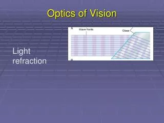
Refraction • This bending of light rays at an angulated interface is known as refraction. • Noteparticularly that the degree of refraction increases as a function of; 1- the ratio ofthe two refractive indices of the two transparent media and 2- the degree of angulationbetween the interface and the entering wave front.
A) Light rays entering a glass surface perpendicular to the light rays B)and a glass surface angulated to the light rays This figuredemonstrates that the distance between waves after they enter the glass is shortened to about two thirds that in air. It also showsthat light rays striking an angulated glass surface are bent.
A- Focusing of light from a point source to a line focus by a cylindricallens. B- Two cylindrical convex lenses at right angles toeach other, demonstrating that one lens converges light rays inone plane and the other lens converges light rays in the plane ata right angle. The two lenses combined give the same point focusas that obtained with a single spherical convex lens.
Two point sources of lightfocused at two separate points onopposite sides of the lens. • B. Formationof an image by a convexspherical lens.
the concave lens divergeslight rays, • the convex lens converges light rays. • Concave cylindrical lenses diverge light rays in onlyone plane in the same manner that convex cylindricallenses converge light rays in one plane.
The more a lens bends light rays, the greater is its“refractive power.” This refractive power is measured interms of diopters. The refractive power in diopters of aconvex lens is equal to 1 meter divided by its focal length.
The refractive power is measured interms of diopters. The refractive power in diopters of aconvex lens is equal to 1 meter divided by its focallength. Thus, a spherical lens that converges parallellight rays to a focal point 1 meter beyond the lens hasarefractive power of +1 diopter If the lens is capable of bending parallel light raystwice as much as a lens with a power of +1 diopter, it issaid to have a strength of +2 diopters, and the light rayscome to a focal point 0.5 meter beyond the lens. A lenscapable of converging parallel light rays to a focal pointonly 10 centimeters (0.10 meter) beyond the lens has arefractive power of +10 diopters.
Optics of Eye • Lens • Pupil • Retina
The eye, is optically equivalentto the usual photographic camera. • It has a lens system,a variable aperture system (the pupil), and a retinathat corresponds to the film.
The lens system of the eyeis composed of four refractive interfaces: 1) Air -Anterior surface of thecornea, 2) Posterior surfaceof the cornea - Aqueous humor, 3) Aqueous humor -Anterior surfaceof the lens of the eye, and 4) Posterior surface of the lens -Vitreoushumor.
The eye as a camera. The numbers are the refractive indices. • The internal index of air is 1; • the cornea, 1.38; • the aqueous humor, 1.33; • the crystalline lens, 1.40; • the vitreous humor, 1.34.
Mechanism of “Accommodation” • In children, the refractive power of the lens of the eyecan be increased voluntarily from 20 diopters to about34 diopters; this in an “accommodation” of 14 diopters. • To do this, the shape of the lens is changed from thatof a moderately convex lens to that of a very convexlens.
When the lens is in a relaxed statewith no tension on its capsule, it assumes an almostspherical shape, owing mainly to the elastic retractionof the lens capsule. about 70 suspensory ligaments attach radiallyaround the lens, pulling the lens edges toward the outer circle of the eyeball These ligaments are constantlytensed by their attachments at the anteriorborder of the choroid and retina. The tension on theligaments causes the lens to remain relatively flatunder normal conditions of the eye.
lateral attachments ofthe lens ligaments to the eyeball is the ciliary muscle; meridional fibers, circular fibers. • Themeridional fibers extend from the peripheral ends ofthe suspensory ligaments to the corneoscleral junction. • When these muscle fibers contract, the peripheralinsertions of the lens ligaments are pulled mediallytoward the edges of the cornea, thereby releasing theligaments’ tension on the lens. • When the circular fibers contract, a sphincterlikeaction occurs, decreasing the diameter of the circleof ligament attachments; this also allows the ligamentsto pull less on the lens capsule.
Thus, contraction of either set of smooth musclefibers in the ciliary muscle relaxes the ligaments to thelens capsule, and • the lens assumes a more sphericalshape, like that of a balloon, because of the naturalelasticity of the lens capsule.
Accommodation Is Controlled by Parasympathetic Nerves • The ciliary muscle is controlled almost entirely byparasympathetic nerve signals transmitted to the eyethrough the third cranial nerve from the third nervenucleus in the brain stem • Stimulation of the parasympathetic nerves contractsboth sets of ciliary muscle fibers, which relaxes the lensligaments, thus allowing the lens to become thickerand increase its refractive power
Presbyopia • As a person grows older, the lens growslarger and thicker and becomes far less elastic, partlybecause of progressive denaturation of the lens proteins. • The ability of the lens to change shape decreaseswith age. • The power of accommodation decreases; İn a child: 14 diopters 45-50 years; 2 diopters 70 years; 0 diopters • Thereafter, the lens remains almost totally nonaccommodating,a condition known aspresbyopia
Emmetropia(Normal Vision) eye is considered to be normal, or “emmetropic,” if parallellight rayia distant objects are in sharp focus onthe retina when the ciliary muscle is completely relaxed. This means that the emmetropic eye can see all distantobjects clearly with its ciliary muscle relaxed. However,to focus objects at close range, the eye must contract itsciliary muscle and thereby provide appropriate degrees of accommodation.
Hyperopia • usually due to either aneyeball that is too short or, occasionally, a lens systemthat is too weak. • In this condition, parallel light rays are not bentsufficiently by the relaxed lens system to come to focusby the time they reach the retina
Myopia when the ciliary muscle is completely relaxed, thelight rays coming from distant objects are focused infront of the retina. This is usually due to too long an eyeball,but it can result from too much refractive power in thelens system of the eye.
Myopia; ? Lens • Hypermetropia; ? Lens
Correction of Myopia and Hyperopia by Use of Lenses. • It will be recalled that light rays passing through a • concave lens diverge. If the refractive surfaces of the eyehave too much refractive power, as in myopia, thisexcessive refractive power can be neutralized by placingin front of the eye a concave spherical lens, which willdiverge rays. • Conversely, in a person who has hyperopia—that is, • someone who has too weak a lens system—the abnormalvision can be corrected by adding refractive powerusing a convex lens in front of the eye.
Astigmatism, demonstrating that light rays focus at one focal distance in one focal plane (plane AC) and at another focal distance in the plane at a right angle (plane BD).
Chart composed of parallel black bars at different angular orientations for determining the axis of astigmatism.
Cataracts “Cataracts” are an especially common eye abnormalitythat occurs mainly in older people.A cataract is a cloudyor opaque area or areas in the lens. In the early stage ofcataract formation, the proteins in some of the lensfibers become denatured. Later, these same proteinscoagulate to form opaque areas in place of the normal transparent protein fibers. When a cataract has obscured light transmission sogreatly that it seriously impairs vision, the condition canbe corrected by surgical removal of the lens.When thisis done, the eye loses a large portion of its refractivepower, which must be replaced by a powerful convexlens in front of the eye; usually, however, an artificialplastic lens is implanted in the eye in place of the removed lens.
Visual Acuity • Theoretically, light from a distant point source, whenfocused on the retina, should be infinitely small. • However, because the lens system of the eye is neverperfect, such a retinal spot ordinarily has a total diameterof about 11 micrometers, even with maximal resolutionof the normal eye optical system. • The spot isbrightest in its center and shades off gradually towardthe edges, as shown by the two-point images.
Clinical Method for Stating Visual Acuity. • The chart fortesting eyes usuallyconsists of letters of different sizesplaced 20 feet away from the person being tested (Snellen). Ifthe person can see well the letters of a size that he orshe should be able to see at 20 feet, the person is saidto have 20/20 vision—that is, normal vision
Determination of Distance of an Object from the Eye— “Depth Perception” A person normally perceives distance by three majormeans: 1) the sizes of the images of known objects on the retina, 2) the phenomenon of moving parallax, 3) the phenomenon of stereopsis. This ability to determine distance is called depth perception.
Perception of distance • by the size of the image on the retinaand • as a result of stereopsis..