Download
1 / 66
2.27k likes | 5.45k Views
Optics of eye. By Dr Farah Amir ALi. The image forming mechanism:. The functioning of eye can be compared with camera. Eyelids : act as shutter of camera Cornea and lens : focusing system of camera Choroid and pigment layer of retina : darkened interior of camera.

E N D
Optics of eye By Dr Farah Amir ALi
The image forming mechanism: • The functioning of eye can be compared with camera. • Eyelids: act as shutter of camera • Cornea and lens: focusing system of camera • Choroid and pigment layer of retina: darkened interior of camera. • Neural retina: film on which images are focused. • Optic nerve and connections: produce sensation of vision
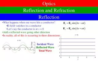
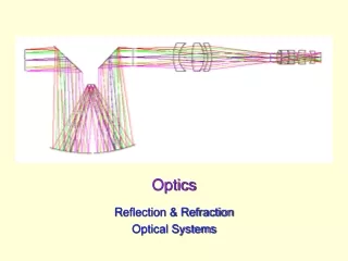
The bending of light rays occur when the ray passes from a medium of one density into medium of a different density. The course of direction of the ray changes if it strikes the surface of new medium at an angle other than perpendicular. Process of refraction
Two factors contribute to the degree of refraction: Comparative densities of two media Angle at which light strikes the 2nd medium Process of refraction
A convex surface curves outward and converge light rays & bring them closer. A concave surface curves inward and diverge light rays Process of refraction
Process of refraction • Parallel light rays striking a biconvex lens are refracted to a point (principal focus) behind the lens. • The principal focus is on a line passing through the centers of curvature of the lens, the principal axis. • The distance between the lens and the principal focus is the principal focal distance..
Practical purposes • For practical purposes, light rays from an object that strike a lens more than 6 m (20 ft) away are considered to be parallel. • The rays from an object closer than 6 m are diverging.
The more a lens bends light rays, the greater is its “refractive power.” This depends on lens curvature. This refractive power is measured in terms of diopters. The refractive power in diopters of a convex lens is equal to 1 meter divided by its focal length. (1/f) Measurement of the Refractive Power of a Lens—“Diopter”
Optics of eye: • The focusing system of eye is composed of several refracting structures which include : the cornea (1.37), the aqueous humour (1.33), the crystalline lens (1.42) and vitrous humour (1.33). • These when combined in action form a very strong refracting system of a short focal length.
Optics of eye: • Reduced eye: • If all the refractive surfaces of the eye are algebraically added together and then considered to be one single lens, the optics of the normal eye may be simplified and represented schematically as a “reduced eye.” This is useful in simple calculations. • The total dioptic power of the eye is about +59 (+60) D at rest out of which about +44 D is contributed by cornea and +16 D by the crystalline lens.
Calculation of 59 diopter Focal point is 17 mm behind lens= 0.017 meter 1/0.017 = 58.8
Accommodation • The process by which curvature of lens is increased. • It is an active process, requiring muscular effort and can therefore be tiring. • Ciliary muscle are the Most Used muscles of body • The nearest point to the eye at which an object can be brought into clear focus by accommodation is called the near point of vision.
Mechanism of accomodation • When the ciliary muscle is relaxed, parallel light rays striking the optically normal (emmetropic) eye are brought to a focus on the retina. • In young individuals, the change in shape may add as many as 12 diopters to the refractive power of the eye.
Accommodation • The near point recedes throughout life, slowly at first and then rapidly with advancing age.(Aging) • NEAR RESPONSE:- • In addition to accommodation, the visual axes converge and the pupil constricts when an individual looks at a near object. This three-part response— accommodation, convergence of the visual axes, and pupillary constriction—is called the near response
Presbyopia: • The near point recedes throughout life, slowly at first and then rapidly with advancing age, • This recession is due principally to increasing hardness of the lens, with a resulting loss of accommodation due to the steady decrease in the degree to which the curvature of the lens can be increased . • Presbyopia is the loss of accomodation by the lens.
Refractive errors: • Emmetropia: refers to normal eye. When ciliary muscle are relaxed, all distant objects are sharp focus on retina.
It is usually due to either an eyeball that is too short or, occasionally, a lens system that is too weak. Hyperopia (Farsightedness)
Hyperopia: • Sustained accommodation, even when viewing distant objects, can partially compensate for the defect, but the prolonged muscular effort is tiring and may cause headaches and blurring of vision. • The prolonged convergence of the visual axes associated with the accommodation may lead eventually to squint. • The defect can be corrected by using glasses with convex lenses, which aid the refractive power of the eye in shortening the focal distance.
In myopia, or “nearsightedness,” when the ciliary muscle is completely relaxed, the light rays coming from distant objects are focused in front of the retina Myopia (Nearsightedness)
Myopia: • Myopia is said to be genetic in origin. • However, there is a positive correlation between sleeping in a lighted room before the age of 2 and the subsequent development of myopia. • In young adult humans the extensive close work involved in activities such as studying accelerates the development of myopia. • This defect can be corrected by glasses with biconcave lenses, which make parallel light rays diverge slightly before they strike the eye
Astigmatism is a refractive error of the eye that causes the visual image in one plane to focus at a different distance from that of the plane at right angles. This most often results from too great a curvature of the cornea in one plane of the eye. Astigmatism
Astigmatism • Astigmatism can usually be corrected with cylindric lenses placed in such a way that they equalize the refraction in all meridians.
Cataracts” are an especially common eye abnormality that occurs mainly in older people. A cataract is a cloudy or opaque area or areas in the lens. In the early stage of cataract formation, the proteins in some of the lens fibers become denatured. Later, these same proteins coagulate to form opaque areas in place of the normal transparent protein fibers Cataracts:
It results from formation of an oddly shaped cornea with prominent bulge on one side causing severe refractive problems. Correction: contact lens Keratoconus
http://www.quantel-medical.com/mediatheque.php?sf=glaucoma • http://www.physpharm.fmd.uwo.ca/undergrad/medsweb/L1Eye/Eye.swf • http://www.physpharm.fmd.uwo.ca/undergrad/medsweb/L1Eye/EyeProb.swf
Pupillary diameter • The major function of the iris is to increase the amount of light that enters the eye during darkness and to decrease the amount of light that enters the eye in daylight. • The pupil of the human eye can become as small as about 1.5 millimeters and as large as 8 millimeters in diameter. • The quantity of light entering the eye can change about 30- fold as a result of changes in pupillary aperture.
“Depth of Focus” of the Lens System Increases with decreasing pupillary diameter?
Depth of Focus:- • The greatest possible depth of focus occurs when the pupil is extremely small. The reason for this is that, with a very small aperture, almost all the rays pass through the center of the lens, and the central most rays are always in focus,
Visual Acuity: • Visual acuity is the degree to which the details and contours of objects are perceived, and it is usually defined in terms of the shortest distance by which two lines can be separated and still be perceived as two lines. • Retinal spot ordinarily has a total diameter of about 11 micrometers, even with maximal resolution of the normal eye optical system.
Visual acuity • A person can normally distinguish two separate points if their centers lie as much as 2 micrometers apart on the retina, which is slightly greater than the width of a foveal cone.
Clinical Method for Stating Visual Acuity. • Clinically, visual acuity is often determined by the use of the familiar Snellen letter charts viewed at a distance of 20 ft (6 m). • The individual being tested reads aloud the smallest line distinguishable. The results are expressed as a fraction. The numerator of the fraction is 20, the distance at which the subject reads the chart. • The denominator is the greatest distance from the chart at which a normal individual can read the smallest line. Normal visual acuity is 20/20
Clinical Method for Stating Visual Acuity. • A subject with 20/15 visual acuity has better than normal vision (not farsightedness); and one with 20/100 visual acuity has subnormal vision.
Depth perception • A person normally perceives distance by three major means: • (1) the sizes of the images of known objects on the retina • (2) the phenomenon of moving parallax, • (3) the phenomenon of stereopsis. (Binocular vision) Note: parallax is the difference in the angular position of two stationary points relative to each other from different viewing positions
Retina: • The retina extends anteriorly almost to the ciliary body. • It is organized in 10 layers and contains the rods and cones, which are the visual receptors, plus four types of neurons: bipolar cells, ganglion cells, horizontal cells, and amacrine cells. • There are many different synaptic transmitters. • The rods and cones, which are next to the choroid, synapse with bipolar cells, and the bipolar cells synapse with ganglion cells.
Retina: • The axons of the ganglion cells converge and leave the eye as the optic nerve. • Horizontal cells connect receptor cells to the other receptor cells in the outer plexiform layer. • Amacrine cells connect ganglion cells to one another in the inner plexiform layer via processes of varying length and patterns.
Rods/cones Rods/cones Horizontal cells Bipolar cells Bipolar cells Amacrine cells Ganglion cell Ganglion cell