Download
1 / 122
1.26k likes | 1.58k Views
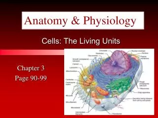
Cells: The Living Units. 3. Cell Theory. The cell is the basic structural and functional unit of life Organismal activity depends on individual and collective activity of cells Biochemical activities of cells are dictated by subcellular structure Continuity of life has a cellular basis.

E N D
Cell Theory • The cell is the basic structural and functional unit of life • Organismal activity depends on individual and collective activity of cells • Biochemical activities of cells are dictated by subcellular structure • Continuity of life has a cellular basis
Structure of a Generalized Cell Figure 3.2
Plasma Membrane • Separates intracellular fluids from extracellular fluids • Plays a dynamic role in cellular activity • Glycocalyx is a glycoprotein area abutting the cell that provides highly specific biological markers by which cells recognize one another
Fluid Mosaic Model • Double bilayer of lipids with imbedded, dispersed proteins • Bilayer consists of phospholipids, cholesterol, and glycolipids • Glycolipids are lipids with bound carbohydrate • Phospholipids have hydrophobic and hydrophilic bipoles
Fluid Mosaic Model Figure 3.3
Functions of Membrane Proteins • Transport of molecules • Enzymatic activity • Receptors for signal transduction Figure 3.4.1
Functions of Membrane Proteins • Intercellular adhesion • Cell-cell recognition • Attachment to cytoskeleton and extracellular matrix Figure 3.4.2
Plasma Membrane Surfaces • Differ in the kind and amount of lipids they contain • Glycolipids are found only in the outer membrane surface • 20% of all membrane lipid is cholesterol (fluidity)
Lipid Rafts • Make up 20% of the outer membrane surface • Composed of sphingolipids and cholesterol • Are concentrating platforms for cell-signaling molecules
Membrane Junctions • Tight junction – impermeable junction that encircles the cell • Desmosome – anchoring junction scattered along the sides of cells • Gap junction – a nexus that allows chemical substances to pass between cells • Together, these promote a coordinated activity of cells by physically binding them together into a cell community
Membrane Junctions: Tight Junction Figure 3.5a
Membrane Junctions: Desmosome Figure 3.5b
Membrane Junctions: Gap Junction Figure 3.5c
Cells: The Living Units Plasma Membrane 3
Passive Membrane Transport: Diffusion • Simple diffusion – nonpolar and lipid-soluble substances • Diffuse directly through the lipid bilayer • Diffuse through channel proteins
Passive Membrane Transport: Diffusion • Facilitated diffusion • Transport of glucose, amino acids, and ions • Transported substances bind carrier proteins or pass through protein channels
Carriers • Are integral transmembrane proteins • Show specificity for certain polar molecules including sugars and amino acids
Diffusion Through the Plasma Membrane Figure 3.7
Passive Membrane Transport: Osmosis • Occurs when the concentration of a solvent is different on opposite sides of a membrane • Diffusion of water across a semipermeable membrane • Osmolarity – total concentration of solute particles in a solution • Tonicity – how a solution affects cell volume
Effect of Membrane Permeability on Diffusion and Osmosis Figure 3.8a
Effect of Membrane Permeability on Diffusion and Osmosis Figure 3.8b
Passive Membrane Transport: Filtration • The passage of water and solutes through a membrane by hydrostatic pressure • Pressure gradient pushes solute-containing fluid from a higher-pressure area to a lower-pressure area
Effects of Solutions of Varying Tonicity • Isotonic – solutions with the same solute concentration as that of the cytosol • Hypertonic – solutions having greater solute concentration than that of the cytosol • Hypotonic – solutions having lesser solute concentration than that of the cytosol
Sodium-Potassium Pump Extracellular fluid K+ is released and Na+ sites are ready to bind Na+ again; the cycle repeats. 6 Binding of cytoplasmic Na+ to the pump protein stimulates phosphorylation by ATP. 1 Cytoplasm Phosphorylation causes the protein to change its shape. 2 Concentration gradients of K+ and Na+ The shape change expels Na+ to the outside, and extracellular K+ binds. 3 Loss of phosphate restores the original conformation of the pump protein. 5 K+ binding triggers release of the phosphate group. 4 Figure 3.10
Active Transport • Uses ATP to move solutes across a membrane • Requires carrier proteins
Types of Active Transport • Symport system – two substances are moved across a membrane in the same direction • Antiport system – two substances are moved across a membrane in opposite directions
Types of Active Transport • Primary active transport – hydrolysis of ATP phosphorylates the transport protein causing conformational change • Secondary active transport – use of an exchange pump (such as the Na+-K+ pump) indirectly to drive the transport of other solutes
Types of Active Transport Figure 3.11
Vesicular Transport • Transport of large particles and macromolecules across plasma membranes • Exocytosis – moves substance from the cell interior to the extracellular space • Endocytosis – enables large particles and macromolecules to enter the cell
Vesicular Transport • Transcytosis – moving substances into, across, and then out of a cell • Vesicular trafficking – moving substances from one area in the cell to another • Phagocytosis – pseudopods engulf solids and bring them into the cell’s interior
Vesicular Transport • Fluid-phase endocytosis – the plasma membrane infolds, bringing extracellular fluid and solutes into the interior of the cell • Receptor-mediated endocytosis – clathrin-coated pits provide the main route for endocytosis and transcytosis • Used by some hormones for cell entry • Non-clathrin-coated vesicles – caveolae that are platforms for a variety of signaling molecules
Exocytosis Figure 3.12a
Clathrin-Mediated Endocytosis Figure 3.13
Membrane Potential • Voltage across a membrane • Resting membrane potential – the point where K+ potential is balanced by the membrane potential • Ranges from –20 to –200 mV • Results from Na+ and K+ concentration gradients across the membrane • Differential permeability of the plasma membrane to Na+ and K+ • Steady state – potential maintained by active transport of ions
Generation and Maintenance of Membrane Potential PLAY InterActive Physiology®: Nervous System I: The Membrane Potential Figure 3.15
Cell Adhesion Molecules (CAMs) • Anchor cells to the extracellular matrix • Assist in movement of cells past one another • Rally protective white blood cells to injured or infected areas
Roles of Membrane Receptors • Contact signaling – important in normal development and immunity • Electrical signaling – voltage-regulated “ion gates” in nerve and muscle tissue • Chemical signaling – neurotransmitters bind to chemically gated channel-linked receptors in nerve and muscle tissue • G protein-linked receptors – ligands bind to a receptor which activates a G protein, causing the release of a second messenger, such as cyclic AMP
Operation of a G Protein • An extracellular ligand (first messenger), binds to a specific plasma membrane protein • The receptor activates a G protein that relays the message to an effector protein
Operation of a G Protein • The effector is an enzyme that produces a second messenger inside the cell • The second messenger activates a kinase • The activated kinase can trigger a variety of cellular responses
Operation of a G Protein Figure 3.16
Cells: The Living Units Cytoplasm 3
Cytoplasm • Cytoplasm – material between plasma membrane and the nucleus • Cytosol – largely water with dissolved protein, salts, sugars, and other solutes • Cytoplasmic organelles – metabolic machinery of the cell • Inclusions – chemical substances such as glycosomes, glycogen granules, and pigment
Cytoplasmic Organelles • Specialized cellular compartments • Membranous • Mitochondria, peroxisomes, lysosomes, endoplasmic reticulum, and Golgi apparatus • Nonmembranous • Cytoskeleton, centrioles, and ribosomes
Mitochondria • Double membrane structure with shelflike cristae • Provide most of the cell’s ATP via aerobic cellular respiration • Contain their own DNA and RNA
Mitochondria Figure 3.17
Ribosomes • Granules containing protein and rRNA • Site of protein synthesis • Free ribosomes synthesize soluble proteins • Membrane-bound ribosomes synthesize proteins to be incorporated into membranes
Endoplasmic Reticulum (ER) • Interconnected tubes and parallel membranes enclosing cisternae • Continuous with the nuclear membrane • Two varieties – rough ER and smooth ER