Download
1 / 61
630 likes | 764 Views
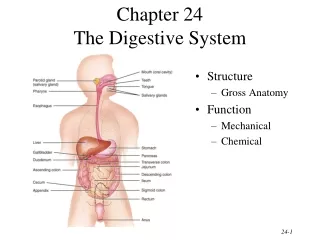
Chapter 24 The Digestive System. BIO 211 Lab Instructor Dr. Gollwitzer. Today in class we will: Discuss the gastrointestinal tract and identify its components Identify the accessory organs associated with the digestive tract

E N D
Chapter 24 The Digestive System BIO 211 Lab Instructor Dr. Gollwitzer
Today in class we will: • Discuss the gastrointestinal tract and identify its components • Identify the accessory organs associated with the digestive tract • The layers that make up the digestive tract wall and each layer’s characteristics • Mucosa • Submucosa • Muscularis externa • Serosa • Begin our discussion of the gastrointestinal tract components in more detail • Oral cavity • Pharynx • Esophagus
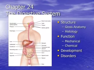
Digestive System • Gastrointestinal (GI) tract = continuous, muscular tube from mouth to anus • Oral cavity (mouth) • Pharynx • Esophagus • Stomach • Small intestine • Large intestine • Anus
Digestive System • Accessory digestive organs = any digestive organ attached to the GI tract • Teeth • Tongue • Glandular organs • Salivary glands • Pancreas • Liver • Gall Bladder
Histological Organization • 4 Major layers to wall • Mucosa • Submucosa • Muscularis externa • Serosa
Mucosa • Inner lining of digestive tract • A mucous membrane • Consists of • Epithelium - moistened by glandular secretions • Lamina propria = areolar CT “filler”
Mucosa: Digestive Epithelia • Depend on location, function, stresses • Simple or stratified • Columnar or squamous
Mucosa: Digestive Epithelia • Nonkeratinized stratified squamous epithelium • Where mechanical stress most severe • In oral cavity, pharynx, esophagus, rectum • Life span = 2-3 days • Simple columnar epithelium • Where absorption and secretion occur (with villi and goblet cells) • Stomach, small intestine, most of large intestine • Life span = 6 days
Mucosa: Lamina Propria • Layer of areolar (loose connective) tissue • Contains • Blood vessels, sensory nerve endings, lymphatic vessels, lymphoid tissue • Mucosal glands and glandular secretions • Muscularis mucosae/interna • Band of smooth muscle and elastic fibers • Smooth muscle arranged in 2 concentric layers • Inner, circular layer (around lumen) • Outer, longitudinal layer • Contractions alter shape of lumen and move epithelial folds
Mucosa: Glandular Structures • Have secretory function • Associated with simple columnar cells and/or mucous-secreting cells • Goblet cells (exocrine) mucus • Enteroendocrine cells • hormones (e.g., G cells gastrin) • Coordinate activities of digestive tract and accessory glands • e.g., chief cells, G cells, parietal cells
Submucosa • Layer of dense, irregular connective tissue • Around muscularis mucosae • Contains • Large blood vessels and lymphatic vessels • Exocrine glands buffers and enzymes into lumen • Submucosal plexus contains nerve fibers and neurons
Muscularis Externa • Dominated by smooth muscle cells • Inner, circular layer • Outer, longitudinal layer • Important role in mechanical processing and movement of materials along tract • Also contains • Lymphoid nodules (Peyer’s patches) • Masses of lymphoid tissue; have lymphocytes that protect small intestine from bacteria that are normal inhabitants of large intestine • Myenteric plexus/plexus of Auerbach • Network of neurons located between circular and longitudinal muscle layers • Movements coordinated by enteric nervous system (part of ANS)
Serosa • A serous membrane = lines sealed, internal subdivisions of ventral body cavity • Covers muscularis externa of most of digestive tract • except oral cavity, pharynx, esophagus, and rectum, which have adventitia (a dense collagen fibrous sheath)
Oral (Buccal) Cavity • Space within mouth • Lined by oral mucosa (nonkeratinized stratified squamous epithelium) • Vestibule = space between cheeks or lips and teeth • Labial frenulum = fold of mucosa; connects lip to gum • Cheeks = lateral walls of oral cavity • Mucosae supported by fat pads and buccinator muscles • Labia (lips) • Gingivae (gums) • Ridges that surround base of each tooth • On alveolar processes of maxillary bones and mandible
Oral (Buccal) Cavity • Palates • Hard palate – formed by maxillary bones (anterior) and palatine bones (posterior) • Soft palate – fleshy part posterior to hard palate • Formed from skeletal muscle • Posterior margin supports uvula = dangling process that helps prevent food from entering pharynx prematurely • Tongue • Muscular organ attached to floor of oral cavity • Dorsum (superior) surface covered with lingual papillae (location of taste buds) • Lingual frenulum – thin fold of mucous membrane that attaches tongue to floor of oral cavity
Pharynx • Throat area posterior to nasal and oral cavities • Shared by respiratory and digestive systems • Extends between internal nares and entrances to larynx and esophagus • Nasopharynx • Posterior portion or nasal cavity • Separated from oral cavity by soft palate • Contains • Pharyngeal tonsils and opening to auditory (eustachian) tube • Oropharynx • Posterior portion of oral cavity • Between soft palate and base of tongue • Fauces = opening between oral cavity and oropharynx • Laryngopharynx • Inferior part • Between hyoid bone and entrances to esophagus and larynx
Esophagus • Hollow, muscular tube • From posterior laryngopharynx to stomach • Descends through thoracic cavity posterior to trachea • Enters abdominopelvic cavity through esophageal hiatus in diaphragm
Esophageal Histology • Muscularis externa transitions from • Skeletal muscle fibers (superior third) to • Smooth muscle fibers (inferior third) • Adventitia • CT outside muscularis externa (no serosa) • Anchors esophagus against dorsal body wall
Esophageal Sphincters • Circular smooth muscles that control entrance/exit to esophagus • Upper esophageal sphincter • In superior 1 in. • Prevents air from entering • Lower esophageal sphincter • At inferior end • Prevents backflow from stomach
Today in class we will: • Complete our more detailed discussion of the gastrointestinal tract components • Stomach • Small intestine • Large intestine • Anus • Begin our discussion of the 4 accessory digestive organs • Teeth • Tongue • Glandular organs • Salivary glands
Stomach • Muscular tube with extra layers of smooth muscle cells • Strengthen stomach wall • Assist in mixing/churning activities required to form chyme • Muscularis mucosae/interna – has extra outer, circular layer • Muscularis externa – has oblique layer
Stomach • Has expanded J shape • Lesser curvature = shorter, inner, medial surface • Greater curvature = longer, outer, lateral surface
Stomach • Has 4 regions • Cardia • Smallest region where esophagus enters (at gastroesophageal/cardiac sphincter) • Fundus • Superior hump to left of cardia that contacts the diaphragm • Body (corpus) • Main (middle) region of the stomach between fundus and curve • Pylorus • Last part of stomach that enters the duodenum through pyloric sphincter
Stomach • Rugae • Folds of gastric mucosa • Temporary features • Let gastric lumen expand (almost disappear when stomach full)
Stomach • Gastric pits • Depressions that open onto gastric surface • Each communicates with several gastric glands • Mucous cells line neck • Gastric glands • In fundus and body of stomach • 2 types of secretory cells • Parietal cells • Chief cells
Small Intestine • 3 Regions • Duodenum (10 in.) • First part; connects to pylorus • “Mixing bowl” • Receives chyme from stomach and secretions from pancreas and liver • Curves in a C that encloses head of the pancreas • Jejunum (8 feet) • Middle part • Site for most of chemical digestion and nutrient absorption • Ileum (12 feet) • Last part • Connects to large intestine via ileocecal valve
Small Intestine • Plicae circulares • inner folds in intestinal lining • Permanent feature -- do not disappear when small intestine fills • Wall • Mucosa • Intestinal epithelium • Simple columnar epithelium; covers villi (pl; singular = villus) • Villus capillaries/villus capillary network • Lacteals • Intestinal glands (goblet cells, enteroendocrine cells) • Muscularis mucosae/interna • Submucosa, muscularis externa, and serosa as described previously
Large Intestine (Bowel) • 3 Main sections • Cecum • Colon (4.5 ft) • Rectum (6 in.)
Large Intestine (Bowel) • Cecum • Pouch-like structure that connects to ileum (at ileocecal valve) • Vermiform appendix = small, worm-like appendage of lymphoid tissue attached to cecum
Large Intestine (Bowel) • Colon • Ascending colon – along right side; takes right turn at superior end = right colic/hepatic flexure • Transverse colon – crosses from R to L; takes right turn downward at left end = left colic/splenic flexure • Descending colon – along left side; curves medially at sigmoid flexure • Sigmoid colon – S-shaped; empties into rectum • Haustra • Pouches caused by longitudinal bands of muscularis externa (taeniae coli) • Permit expansion and elongation of colon
Large Intestine (Bowel) • Rectum • Anal canal = last portion • Contain small longitudinal folds (anal columns) • Anus/anal orifice = exit of anal canal • Contains 2 sphincters • Internal anal sphincter – smooth muscle, involuntary control • External anal sphincter – skeletal muscle, voluntary control
Teeth • Located in alveoli (bony sockets) on alveolar processes of maxillary bones and mandible • Number • Primary (deciduous) • 5/side/jaw = 20 • Secondary • +3 molars/side/jaw = 32 • Replace primary teeth
Teeth • Parts • Crown – above gum line • Neck – boundary between crown and root • Root – below gum line; sits in alveolus
Teeth • Layers/Regions • Outer • Enamel • Thin, white layer that covers crown • Hardest biologically manufactured substance • Cementum • Thin layer that covers root; protects and helps anchor tooth • Inner • Dentin • Makes up bulk of tooth • Mineralized matrix (CaPO4 crystals) similar to bone, except acellular • Pulp cavity • Central region • Receives blood vessels and nerves from root canal • Root canal • Narrow tunnel at root/base of tooth • Blood vessels and nerves enter through apical foramen
Salivary Glands • Outside oral cavity, but secrete into oral cavity • 3 pairs • Parotid glands • Extend from mastoid process of temporal bone across outer surface of masseter muscle • Parotid (Stenson’s) duct – from parotid through buccinator to oral cavity • Sublingual glands • Under the floor of the mouth • Many small sublingual (Rivinus’) ducts open along side of lingual frenulum • Submandibular glands • Along inner surfaces of mandible • Submandibular (Wharton’s) ducts – open into mouth on either side of lingual frenulum immediately posterior to teeth