Download
1 / 41
420 likes | 445 Views
Chapter 15 Sense Organs. Sensory Receptors. Sensory receptors make it possible for the body to respond to stimuli caused by changes occurring in the internal or external environment Receptor response General function—responds to stimuli by converting them to nerve impulses

E N D
Sensory Receptors • Sensory receptors make it possible for the body to respond to stimuli caused by changes occurring in the internal or external environment • Receptor response • General function—responds to stimuli by converting them to nerve impulses • Different types of receptors respond to different stimuli • Receptor potential • Develops when an adequate stimulus acts on a receptor; is a graded response • When a threshold is reached, an action potential in the sensory neuron’s axon is triggered • Impulses travel over sensory pathways to the brain and spinal cord, where either they are interpreted as a particular sensation or they initiate a reflex action • Adaptation—a functional characteristic of receptors; receptor potential decreases over time in response to a continuous stimulus, which leads to a decreased rate of impulse conduction and a decreased intensity of sensation
Sensory Receptors • Distribution of receptors • Receptors for special senses of smell, taste, vision, hearing, and equilibrium are grouped into localized areas or into complex organs • General sense organs of somatic senses are microscopic receptors widely distributed throughout the body in skin, mucosa, connective tissue, muscles, tendons, joints, and viscera
Sensory Receptors • Classification of receptors by the following: • Location • Type of stimulus that causes response
Sensory Receptors • Classification by location • Exteroceptors • On or near body surface • Often called cutaneous receptors; examples are pressure, touch, pain, and temperature • Visceroceptors (interoceptors) • Located internally—often within body organs, or viscera • Provide the body with information about internal environment; examples are pressure, stretch, chemical changes, and hunger and thirst • Proprioceptors: specialized type of visceroceptor • Location limited to skeletal muscle, joint capsules, and tendons • Provide information on body movement, orientation in space, and muscle stretch • Two types—tonic and phasic receptors provide positional information on the body or body parts while at rest or during movement
Sensory Receptors • Classification by stimulus detected • Mechanoreceptors—activated when “deformed” to generate receptor potential • Chemoreceptors—activated by amount or changing concentration of certain chemicals; e.g., taste and smell • Thermoreceptors—activated by changes in temperature • Nociceptors—activated by intense stimuli that may damage tissue; the sensation produced is pain • Photoreceptors—found only in the eye; respond to light stimuli if the intensity is great enough to generate a receptor potential • Osmoreceptors—concentrated in the hypothalamus; activated by changes in concentration of electrolytes (osmolarity) in extracellular fluids
Sensory Receptors • Classification by structure (Figure 15-2)—divides sensory receptors into either those with free nerve endings or those with encapsulated nerve endings • Free nerve endings • Most widely distributed type of sensory receptor • Include both exteroceptors and visceroceptors • Called nociceptors—are primary receptors for pain • Other sensations mediated include itching, tickling, touch, movement, and mechanical stretching • Primary receptors for heat and cold • Two types of nerve fibers carry pain impulses from nociceptors to the brain: • Acute (A) fibers—mediate sharp, intense, localized pain • Chronic (B) fibers—mediate less intense, but more persistent, dull or aching pain
Sensory Receptors • Classification by structure (cont.) • Other free nerve ending receptors • Root hair plexuses • Weblike arrangements of free nerve endings around hair follicles • Merkel discs • Mediate sensations of discriminative touch
Sensory Receptors • Stretch receptors—two types; muscle spindles and Golgi tendon receptors operate to provide body with information concerning muscle length and strength of muscle contraction • Muscle spindle—composed of 5 to 10 intrafusal fibers lying between and parallel to regular (extrafusal) muscle fibers • Large diameter and rapid conducting, type Ia, and smaller diameter and slower conducting, type II, afferent fibers carry messages to brain concerning changes in muscle length • If length of a muscle exceeds a certain limit, a stretch reflex is initiated to shorten the muscle, thus helping to maintain posture • Golgi tendon organs—located at junction between muscle tissue and tendon (Figure 15-2) • Type Ib sensory neurons are stimulated by excessive contraction—when stimulated, they cause muscle to relax • Golgi tendon reflex protects muscle from tearing internally as a result of excessive contractile force
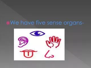
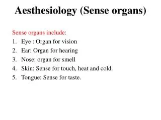
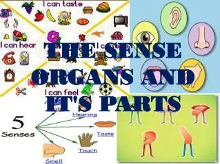
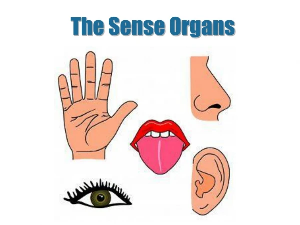
Special Senses • Characterized by receptors grouped closely together or grouped in specialized organs; senses of smell, taste, hearing, equilibrium, and vision
Sense of Smell • Olfactory receptors • Olfactory sense organs consist of epithelial support cells and specialized olfactory receptor neurons (Figure 15-4) • Olfactory cilia—located on olfactory receptor neurons that touch the olfactory epithelium lining the upper surface of nasal cavity • Olfactory cells—chemoreceptors; gas molecules or chemicals dissolved in mucus covering the nasal epithelium stimulate olfactory cells • Olfactory epithelium—located in most superior portion of nasal cavity • Olfactory receptors—extremely sensitive and easily fatigued
Sense of Smell • Olfactory pathway—when level of odor-producing chemicals reaches a threshold level, the following occurs (Figure 15-5): • Receptor potential, and then action potential, is generated and passed to the olfactory nerves in the olfactory bulb • The impulse then passes through the olfactory tract and into the thalamic and olfactory centers of brain for interpretation, integration, and memory storage
Sense of Taste • Taste buds—sense organs that respond to gustatory, or taste, stimuli; associated with papillae • Chemoreceptors that are stimulated by chemicals dissolved in the saliva • Gustatory cells—specialized cells in taste buds; gustatory hairs extend from each gustatory cell into the taste pore • Sense of taste depends on the creation of a receptor potential in gustatory cells as a result of taste-producing chemicals in the saliva • Taste buds are similar structurally; functionally, each taste bud responds most effectively to one of four primary taste sensations: sour, sweet, bitter, and salty (and perhaps metallic and umami) (Figure 15-6) • Adaptation and sensitivity thresholds are different for each of the primary taste sensations
Sense of Taste • Neuronal pathway for taste • Taste sensation begins with a receptor potential in gustatory cells of a taste bud; generation and propagation of an action potential then transmits sensory input to the brain • Nerve impulses from anterior two thirds of the tongue travel over the facial nerve; those from posterior one third of the tongue travel over the glossopharyngeal nerve; vagus nerve plays a minor role in taste • Nerve impulses are carried to the medulla oblongata, relayed into the thalamus, and then into the gustatory area of the cerebral cortex in the parietal lobe of the brain
Sense of Hearing and Balance: The Ear • External ear—two divisions (Figures 15-7 and 15-8): • Auricle, or pinna—visible portion of the ear • External auditory meatus—tube leading from auricle into the temporal bone and ending at the tympanic membrane
Sense of Hearing and Balance: The Ear • Middle ear (Figure 15-8) • Tiny, epithelium-lined cavity hollowed out of the temporal bone • Contains three auditory ossicles • Malleus (hammer)—attached to inner surface of tympanic membrane • Incus (anvil)—attached to malleus and stapes • Stapes (stirrup)—attached to incus • Openings into middle ear cavity • Opening from external auditory meatus covered with tympanic membrane • Oval window—opening into inner ear; stapes fits here • Round window—opening into inner ear; covered by a membrane • Opening into the auditory (eustachian) tube
Sense of Hearing and Balance: The Ear • Inner ear (Figure 15-9, A) • Structure of the inner ear • Bony labyrinth—made up of the vestibule, the cochlea, and semicircular canals • Membranous labyrinth—made up of utricle and saccule inside the vestibule, cochlear duct inside the cochlea, and the membranous semicircular canals inside the bony ones • Vestibule and semicircular canals are involved with balance • Cochlea—involved with hearing • Endolymph—clear, potassium-rich fluid filling the membranous labyrinth • Perilymph—similar to cerebrospinal fluid, surrounds the membranous labyrinth, filling space between the membranous tunnel and its contents and the bony walls that surround it
Sense of Hearing and Balance: The Ear • Inner ear (cont.) • Cochlea and cochlear duct (Figure 15-9, B) • Cochlea—bony labyrinth • Modiolus—cone-shaped core of the bone that houses the spiral ganglion, which consists of cell bodies of the first sensory neurons in the auditory relay • Cochlear duct • Lies inside the cochlea; only part of the internal ear concerned with hearing; contains endolymph • Shaped like a triangular tube • Divides cochlea into the scala vestibuli, the upper section, and the scala tympani, the lower section; both sections filled with perilymph • Vestibular membrane—roof of cochlear duct • Basilar membrane—floor of cochlear duct • Organ of Corti—rests on basilar membrane; consists of supporting cells and hair cells • Axons of the neurons that begin around the organ of Corti, extend in the cochlear nerve to the brain to produce the sensation of hearing
Sense of Hearing and Balance: The Ear • Inner ear (cont.) • Sense of hearing • Sound is created by vibrations • Ability to hear sound waves depends on volume, pitch, and other acoustic properties • Sound waves must be of sufficient amplitude to move the tympanic membrane and have a frequency capable of stimulating the hair cells in the organ of Corti • Basilar membrane is not the same width and thickness throughout its length; high-frequency sound waves vibrate the narrow portion near the oval window, whereas low frequencies vibrate the wider, thicker portion near the apex of the cochlea; this fact allows different hair cells to be stimulated and different pitches of sound to be perceived • Perception of loudness is determined by the amplitude of the movement of basilar membrane; the greater the movement, the louder the perceived sound • Hearing—results from stimulation of auditory area of cerebral cortex
Sense of Hearing and Balance: The Ear • Sense of hearing (cont.) • Pathway of sound waves (Figure 15-10) • Enter external auditory canal • Strike tympanic membrane, causing vibrations • Tympanic vibrations move malleus, which in turn moves incus and then stapes • Stapes moves against oval window, which begins fluid conduction of sound waves • The perilymph in the scala vestibuli of cochlea begins “ripple” that is transmitted through vestibular membrane to endolymph inside duct, to basilar membrane, then to organ of Corti • From basilar membrane, ripple is transmitted through perilymph in scala tympani and then expends itself against round window
Sense of Hearing and Balance: The Ear • Sense of hearing (cont.) • Neuronal pathway of hearing • A movement of the hair cells against tectorial membrane stimulates dendrites that terminate around base of hair cells and initiates impulse conduction by the cochlear nerve to the brainstem • Impulses pass through “relay stations” in the nuclei in medulla, pons, midbrain, and thalamus before reaching auditory area of temporal lobe
Sense of Hearing and Balance: The Ear • Vestibule and semicircular canals (Figure 15-9, A) • Vestibule—the central section of the bony labyrinth; the utricle and saccule are the membranous structures within the vestibule • Semicircular canals—three, each at right angles to the others, are found in each temporal bone; within the bony semicircular canals are the membranous semicircular canals, each containing endolymph and connecting with the utricle; near this junction, each canal enlarges into an ampulla
Sense of Hearing and Balance: The Ear • Vestibule and semicircular canals (cont.) • Sense of balance • Static equilibrium—ability to sense the position of the head relative to gravity or to sense acceleration or deceleration (Figure 15-11) • Movements of the macula, located in both the utricle and saccule almost at right angles to each other, provide information related to head position or acceleration • Otoliths are located within matrix of macula • Changing head position produces a change of pressure on the otolith-weighted matrix, which stimulates hair cells that, in turn, stimulate receptors of vestibular nerve • Vestibular nerve fibers conduct impulses to the brain and produce a sensation of the position of the head and also a sensation of a change in the pull of gravity • Righting reflexes—muscular responses to restore the body and its parts to their normal position when they have been displaced; caused by stimuli of macula and impulses from proprioceptors and from eyes
Sense of Hearing and Balance: The Ear • Sense of balance (cont.) • Dynamic equilibrium—needed to maintain balance when head or body is rotated or suddenly moved; able to detect changes both in direction and rate at which movement occurs (Figure 15-12) • Depends on functioning of cristae ampullaris, which are located in ampulla of each semicircular canal • Cupula—gelatinous cap in which hair cells of each crista are embedded; does not respond to gravity; moves with flow of endolymph in semicircular canals • Semicircular canals are placed at almost right angles to each other to detect movement in all directions • When the cupula moves, hair cells are bent, producing a receptor potential followed by an action potential; action potential passes through vestibular portion of eighth cranial nerve to medulla oblongata, where it is sent to other areas of brain and spinal cord for interpretation, integration, and response
Vision: The Eye • Structure of the eye (Figures 15-13 and 15-14) • Coats of the eyeball—three layers of tissues compose the eyeball: • Sclera—outer coat • Tough, white, fibrous tissue • Cornea—transparent anterior portion that lies over iris; no blood vessels found in cornea or in lens • Canal of Schlemm—ring-shaped venous sinus found deep within anterior portion of the sclera at its junction with the cornea • Choroid—middle coat • Contains many blood vessels and a large amount of pigment • Anterior portion has three different structures (Figure 15-14): • Ciliary body—thickening of choroid, fits between anterior margin of retina and posterior margin of iris; ciliary muscle lies in anterior part of ciliary body; ciliary processes—fold in ciliary body • Suspensory ligament—attached to ciliary processes and blends with elastic capsule of the lens, to hold it in place • Iris—colored part of eye; consists of circular and radial smooth muscle fibers that form a doughnut-shaped structure; attaches to ciliary body
Vision: The Eye • Coats of the eyeball (cont.) • Retina—incomplete innermost coat of eyeball • Three layers of neurons make up the sensory retina (Figure 15-16): • Photoreceptor neurons—visual receptors, highly specialized for stimulation by light rays • Rods—absent from fovea and macula; increased in density toward periphery of retina • Cones—less numerous than rods; most densely concentrated in fovea centralis in macula lutea • Bipolar neurons • Ganglionic neurons—all axons of these neurons extend back to the optic disc; part of sclera, which contains perforations through which the fibers emerge from the eyeball as the optic nerve
Vision: The Eye • Cavities and humors • Cavities—eyeball has a large interior space divided into two cavities: • Anterior cavity—lies in front of lens; has two subdivisions • Anterior chamber—space anterior to iris and posterior to cornea • Posterior chamber—small space posterior to iris and anterior to lens • Posterior cavity—larger than anterior cavity; occupies all the space posterior to lens, suspensory ligament, and ciliary body
Vision: The Eye • Cavities and humors (cont.) • Humors • Aqueous humor—fills both chambers of anterior cavity; clear, watery fluid that often leaks out when eye is injured; formed from blood in capillaries located in ciliary body (Figure 15-17) • Vitreous humor—fills posterior cavity; semisolid material; helps to maintain sufficient intraocular pressure, with aqueous humor, to give the eyeball its shape
Vision: The Eye • Muscles—two types of eye muscles: • Extrinsic eye muscles (Figure 15-18)—skeletal muscles that attach to the outside of the eyeball and to the bones of the orbit; named according to their position on eyeball; the muscles are superior, inferior, medial, and lateral rectus muscles and superior and inferior oblique muscles • Intrinsic eye muscles—smooth muscles located within the eye: iris—regulates size of pupil; ciliary muscle—controls shape of lens
Vision: The Eye • Accessory structures (Figure 15-19) • Eyebrows and eyelashes—give some protection against foreign objects entering eye; cosmetic purposes • Eyelids—consist of voluntary muscle and skin with a tarsal plate; lined with conjunctiva, a mucous membrane; palpebral fissure—opening between the eyelids; canthus—where upper and lower eyelids join • Lacrimal apparatus—structures that secrete tears and drain them from surface of eyeball (Figure 15-21) • Lacrimal glands—size and shape of a small almond; located at upper, outer margin of each orbit; approximately a dozen small ducts lead from each gland; drain tears onto conjunctiva • Lacrimal canals—small channels that empty into lacrimal sacs • Lacrimal sacs—located in a groove in lacrimal bone • Nasolacrimal ducts—small tubes that extend from lacrimal sac into inferior meatus of nose
Vision: The Eye • The process of seeing • Formation of retinal image • Refraction of light rays—deflection, or bending, of light rays produced by light rays passing obliquely from one transparent medium into another of different optical density; cornea, aqueous humor, lens, and vitreous humor are the refracting media of the eye • Accommodation of the lens—increase in curvature of lens to achieve the greater refraction needed for near vision (Figure 15-22) • Constriction of the pupil—muscles of iris are important to formation of a clear retinal image; pupil constriction prevents divergent rays from object from entering eye through periphery of the cornea and lens; near reflex—constriction of pupil that occurs with accommodation of lens in near vision; photopupil reflex—pupil constricts in bright light • Convergence of the eyes—movement of the two eyeballs inward so that their visual axes come together at the object viewed; the closer the object, the greater the degree of convergence necessary to maintain single vision; for convergence to occur, a functional balance between antagonistic extrinsic muscles must exist
Vision: The Eye • The process of seeing (cont.) • Role of photopigments—light-sensitive pigmented compounds undergo structural changes that result in generation of nerve impulses, which are interpreted by the brain as sight • Rods—photopigment in rods is rhodopsin; highly light-sensitive; breaks down into opsin and retinal; separation of opsin and retinal in the presence of light causes an action potential in rod cells; energy is needed to reform rhodopsin (Figure 15-24) • Cones—three types of cones are present in retina, with each having a different photopigment; cone pigments are less light-sensitive than rhodopsin and need brighter light to break down
Vision: The Eye • The process of seeing (cont.) • Neuronal pathway of vision (Figure 15-25) • Fibers that conduct impulses from rods and cones reach the visual cortex in occipital lobes via optic nerves, optic chiasma, optic tracts, and optic radiations • Optic nerve contains fibers from only one retina, but optic chiasma contains fibers from the nasal portion of both retinas; these anatomical facts explain peculiar visual abnormalities that sometimes occur
Cycle of Life: Sense Organs • Sensory information is acquired through depolarization of sensory nerve endings • Age, disease, structural defects, or lack of maturation affect ability to identify and respond • Structure and function response capabilities are related to developmental factors associated with age
Cycle of Life: Sense Organs • Senses become more acute with maturation • Late adulthood—loss of sensory capability • Structural change in receptor cells or other sense organ structures