Download
1 / 103
1.1k likes | 1.6k Views
Function of the Sense Organs. What will we discuss in this chapter? (Outline). Ⅰ. Receptor 1. concepts and classification 2. general properties of receptors Ⅱ. Visual Sense Organ 1. dioptric system 2. light perception and signal processing in the retina

E N D
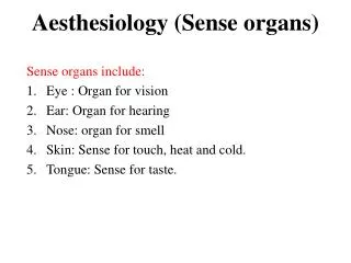
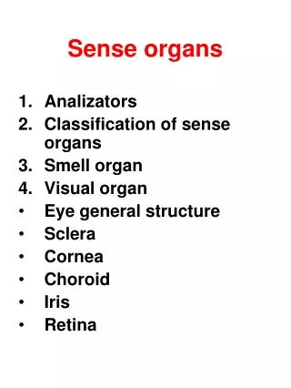
What will we discuss in this chapter?(Outline) Ⅰ. Receptor 1. concepts and classification 2. general properties of receptors Ⅱ. Visual Sense Organ 1. dioptric system 2. light perception and signal processing in the retina 3. vision related terminology Ⅲ. Hearing 1. external ear and middle ear 2. inner ear (cochlea) 3. AP in auditory nerve Ⅳ. Vestibular apparatus 1. receptor 2. adequate stimulus 3. vestibular reaction Ⅴ. Other receptors (sense of smell and sense of taste)
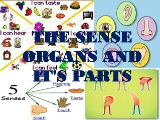
Introduction • Human life would be very different without the ability to sense and perceive external stimulus. • Imagine your world without the ability to see, hear, smell, touch, and feel……
Ⅰ. Receptor Which receptor?
⒈ Concepts and Classification(1) Concepts • Receptor is referred to the organ or structure located on the body surface or within tissues, the function of which is to detect the changes in internal or external environments and to convert stimulus into electrical signals. • In a word: • A state of awareness of a stimulus. • A part of neuron or a specialized cell.
(2) Classification(A) • Location 1) Exteroceptors Located on the body surface or specialized to detect external stimuli Pressure, pain, temp, touch, etc. 2) Visceral receptors Located within internal organs, detect internal stimuli, Blood pressure, pain, fullness. 3) Proprioceptors Found in the joints and muscles, Also in the vestibular structures and the semicircular canals of the inner ear. Limb and body position and movement.
(2) Classification(B) • Modalities 1) Mechanoreceptor Detects stimuli which mechanically deform the receptor; pressure, vibration, touch, sound. 2) Thermoreceptor Detects changes in temperature, hot/cold; 3) Nociceptor (pain) Detects damage to the structures; 4) Photoreceptor Detect light, vision, retina of the eye; 5) Chemoreceptor Detect chemical stimuli; CO2 and O2 in the blood, glucose, small, taste.
(2) Classification(C) • Complexity 1) Simple receptors Usually a single modified dendrite General sense; Touch, pressure, pain, vibration, temperature; 2) Complex receptors High modified dendrites, organized into complex structures; ear, eye. Special senses: Vision, hearing, smell, taste.
2. General properties of receptors • Adequate stimulus of sensory receptors • Transducer function of sensory receptors • Encoding of sensory receptor • Adaptation of sensory receptor
Summary • The external & internal environments are monitored by sensory receptors. • Each type of receptor is excited most effectively by only one modality of stimulus known as the adequate stimulus. • The stimulus is converted into an electrical potential. • Stimuli are detected as either static or dynamic events. • The intensity & duration of the stimulus is frequency coded as bursts of action potentials in the primary afferent nerve.
Eye functions like a camera • Iris allows light into eye • Cornea, Lens & humors focus light onto retina • Light striking retina is converted into action potentials relayed to brain. Functions of the Complete Eye
A slightly irregular hollow sphere with anterior and posterior poles; • The wall is composed of three tunics – fibrous, vascular, and sensory; • The internal cavity is filled with fluids called humors; • The lens separates the internal cavity into anterior and posterior segments. • Dioptric system:cornea, humors, lens, vitreous chamber.
(2) Reduced eye * • Reduced eye is an artificial model. • Calculation of image: Optical Parameter: anteroposterior diameter: 20 mm refractive index : 1.333 radius of curvature : 5 mm AB ab = Bn nb
(3) Accommodation of eye * • Accommodation of lens • near point: • far point: • Pupillary reflex • Decrease size of pupils (parasympathetic) prevents divergent light rays from entering • near reflex of the pupil • pupillary light reflex • Convergence of eyeballs • Viewing near object causes reflexly both eyes to move inward to focus on a near object, this process is called convergence reflex.
(4) Error of refraction * • Error of refraction :Caused by shape of eye and/or power of lens • Myopia • Hyperopia • Astigmatism • Presbyopia • Definition:The crystalline lens tends to harden and the capsule itself becomes less elastic with age • Treatment :convex lens
Emmetropia Myopia Hyperopia Astigmatism Ametropia and Correction
⒉ Light perception and signal processing in the retina( 1) Structure of Retina From outside to inside: Pigment cell Photosensory cell Bipolar cell Ganglion cell rods cones
(2) Photosensory cell (A) • The photoreceptor cells are two types, rod cells (rods) and cone cells (cones)
(2) Photosensory cell (B) • The outer segment of a rod cell has a rod-like appearance, whereas that of a cone cell has a cone-shaped appearance. • The outer segments of the photoreceptor cell contain stacks of membranous discs. • The visual pigments appear to be built into the disc membranes.
(2) Photosensory cell (C) • Distribution of the cones and rods on the retina.
(2) Photosensory cell (D) • Characteristics • Cones see detail but require bright light • Rods see in low light but lack detail
located mainly in fovea; • work best in bright light; • enable us to see fine detail; • responsible for color vision; • each cone has its own bipolar and ganglion cell; • this allows us to see detail but bright light is needed. rods Comparison cones • located mainly in periphery of retina; • responsible for night vision; • detail not detected; • see black, white, and gray (no color); • several rods share 1 bipolar and 1 ganglion cell; • rod vision lacks detail, but, by combining their efforts, groups of rods allow us to see in low light.
cones multicolor rods Black and white
(3) Two types of retinal transduction systems • Other evidence that two photoreceptor system of retina exit. • The nocturnal animals have a preponderance of rods, whereas the diurnal animals have a preponderance of cones in their retina. • The visual pigment in the rods is only rhodopsin. There are three classes of cones in the retina, each containing different pigment sensitive to particular region of visible spectrum.
(4) General transduction mechanism of rods • Photopigments are located in the membrane of the outer segment of rods and cones; • Each pigment consists of an opsin (a protein) and retinal (a lipid); • In the dark, membrane Na+ channels are open -> glutamate is released which depolarizes the membrane • Light splits the opsin and retinal apart-> • Activates transduction (G protein)-> • Activates phosphodiesterase-> • Reduces cGMP -> closes Na+ channels • The net effect of light is to hyperpolarize the retinal receptor and reduce the release of glutamate.
Trichromatic Theory of Color Vision Light of a single wavelength
Trichromatic theory • Occurs at the receptor level ; • Each cone is coated by one of 3 photopigments: • Short-wave (blue) • Medium-wave (green) • Long-wave (red) • Ratio of activated cones = color differentiation; • Primary Colors:sets of 3 colors that can be mixed to produce any other color; • For Visual System:set of interest is “Red Green and Blue”. Theories of Color Vision Young Helmholtz
Color Blindness • Sex-linked conditions: genes on X chromosome, so more common in men. • Protanopia, missing red photopigment; • Deuteranopia : missing green photopigment; • Non-sex-linked condition: • Tritanopia , missing blue photopigment or blue cones • Monochromats : people who are totally colorblind, more severe.
Color Vision Systems Tritanopia deuteranopia protanopia
Blind point (盲点) :In the visual field of each eye, there is a physiological scotoma, the blind point, which coincides with the place where the optic nerve passes out of the eye through the sclera and there is no retina.
3. Vision related terminology • Visual acuity • Visual field • Dark adaptation • Light adaptation • After image • Fusion phenomenon • Stereopsis
Visual Acuity • Visual acuityis defined as the ratio of the distance of the individual from the chart to the distance at which the details of the correctly read line subtend 1'of arc. • Visual angle : • Visual chart
Dark Adaptation and Light Adaptation • Dark adaptation • Definition:On going from a light environment into a darker one, there is a gradual increase in sensitivity allowing dimmer lights to be seen, a mechanism known as dark adaptation. • Mechanism: • Light adaptation • Definition:When one passes suddenly from a dim to a brightly lighted environment, the light seems intensely and even uncomfortably bright until the eyes adapt to the increased illumination and the visual threshold rise. This adaptation occurs over a period of seconds. • Mechanism:
Visual field • The field of the view that can be seen without moving the head is known as the visual field. • white > blue > red> green • narrow:wide: • After image • Fusion phenomenon • Binocular vision • Stereopsis
Hearing Threshold • Hearing thresholdis the lowest intensity that the faintest sound could be heard. • Maximum hearing threshold • Audible area
Properties of Sound • Sound travels in waves as does light • 1. Pitch:determined by “frequency,” the number of cycles per second of a sound wave, measured in hertz (Hz). • 2. Loudness:determined by “amplitude” (height) of the sound wave, measured in decibels (dB) . • 3. Timbre:determined by “complexity and shape” of the sound wave, gives each sound its unique quality.
pitch frequency (Hz) loudness amplitude(dB)
0 dB = hearing threshold • 50 dB = normal conversation • 90 dB = danger zone • 120 dB = rock concert • 130 dB = pain threshold Loudness of Sound