Download
1 / 135
1.43k likes | 1.85k Views
Principles of Neoplasia And Growth Disorders. Dr: Wael H Mansy, MD Assistant Professor Clinical Pharmacy Department-College of Pharmacy King Saud University. Study objectives. • List the main characteristics of benign and malignant tumors.

E N D
Principles of Neoplasia And Growth Disorders Dr: Wael H Mansy, MD Assistant Professor Clinical Pharmacy Department-College of Pharmacy King Saud University
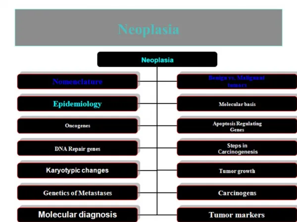
Study objectives • List the main characteristics of benign and malignant tumors. • Describe the nomenclature used for various types of tumors. • Discuss tumor metastasis. How do tumors facilitate their own spread? What are some common sites of metastasis for tumors? • Define oncogenesis. Discuss some of the theories of how it might occur. List some viruses that are oncogenic, as well as the cancers they may cause.
• What is a carcinogen? List some substances that are carcinogenic. • List the local and systemic effects of cancer. • Define cancer cachexia. Why might it occur? • Describe the system by which tumors are “staged.” • What are tumor cell markers? How are they used clinically? • Discuss the various treatment options for cancer. Include the draw-backs of each.
The term neoplasmrefers to an abnormal mass of tissue in which the growth exceeds and is uncoordinated with that of the normal tissues. • Unlike normal cellular adaptive processes such as hypertrophy and hyperplasia, neoplasms do not obey the laws of normal cell growth. • They serve no useful purpose, they do not occur in response to an appropriate stimulus and they continue to grow at the expense of the host.
Cell Cycle • The process of cell growth and division is called the cell cycle. • It is divided into four phases: 1-G1, the postmitotic phase, during which DNA synthesis ceases while RNA and protein synthesis and cell growth take place; 2-S the phase during which DNA synthesis occurs, giving rise to two separate sets of chromosomes;
3-G2 phase: the premitotic phase, during which RNA and protein synthesis continues; 4-M: the phase of cell mitosis or cell division. 5-G0 phase: is a resting or quiescent phase in which nondividing cells reside. • The entry into and progression through the various stages of the cell cycle are controlled by cyclins, cyclin-dependent kinases, and cyclin-dependent kinase inhibitors
Normal tissue renewal and repair involves cell proliferation, differentiation and apoptosis. • Proliferation, or the process of cell division, is an inherent adaptive mechanism for cell replacement when old cells die or additional cells are needed. • Differentiation is the process of specialization whereby new cells acquire the structure and function of the cells they replace. • Apoptosis is a form of programmed cell death that eliminates senescent cells, cells with damaged DNA, or unwanted cells.
Body cells can be divided into two large groups: the well differentiated neurons and cells of skeletal and cardiac muscle that rarely divide and reproduce. • The progenitor or parent cells that continue to divide and reproduce, such as blood cells, skin cells, and liver cells. • A third category of cells are the stem cells that remain quiescent until there is a need for cell replenishment, in which case they divide, producing other stem cells and cells that can carry out the functions of differentiated cells.
Stem cells have two important properties, those of self-renewal and potency. • Self-renewal means that the stem cells can undergo numerous mitotic divisions while maintaining an undifferentiated state. • The term potency is used to define the differentiation potential of stem cells.
A neoplasm, benign or malignant, represents a new growth. ■ Benign neoplasms are well-differentiated tumors that resemble the tissues of origin but have lost the ability to control cell proliferation. They grow by expansion, are enclosed in a fibrous capsule, and do not cause death unless their location is such that it interrupts vital body functions.
■ Malignant neoplasms are less well-differentiated tumors that have lost the ability to control both cell proliferation and differentiation. They grow in a disorganized and uncontrolled manner to invade surrounding tissues, have cells that break loose and travel to distant sites to form metastases, and inevitably cause suffering and death unless their growth can be controlled through treatment.
Cancer Cell Characteristics • Cancer cells are characterized by two main features: (1) abnormal and rapid proliferation; and (2) loss of differentiation so that they do not exhibit normal features and properties of differentiated cells, and hence are more similar to embryonic cells.
ANAPLASIA • Means loss of cell differentiation in cancerous tissue. • In undifferentiated cancer cells both the cells and nuclei display variations in size and shape, a condition referred to as pleomorphism. • Their nuclei are variable in size and bizarre in shape, their chromatin is coarse and clumped, and their nucleoli are often considerably larger than normal.
Some cancers display only slight anaplasia, whereas others display marked anaplasia. • The cytologic/histologic grading of tumors is based on the degree of differentiation and the number of proliferating cells. • The closer the tumor cells resemble comparable normal tissue cells, both morphologically and functionally, the lower the grade. • Accordingly, on a scale ranging from grades I to IV, grade I neoplasms are well differentiated and grade IV are poorly differentiated and display marked anaplasia.
1-Genetic Instability. • Most cancer cells exhibit a characteristic called genetic instability that is often considered to be a hallmark of cancer. • It is thought that cancer cells have a “mutation phenotype” with genetic instability that contributes to the development and progression of cancer.
Characteristics of genetic instability include aneuploidy, in which chromosomes are lost or gained; intrachromosomal instability, which includes insertions, deletions, and amplifications; microsatellite instability, which involves short, repetitive sequences of DNA; and point mutations.
2-Growth Factor Independence • *The ability to proliferate even in the absence of growth factors. This characteristic is often observed when cancer cells are propagated in cell culture. • *The addition of serum, which is rich in growth factors, is unnecessary for the cancers to proliferate. • *Breast cancer cells that do not express estrogen receptors are an example. These cancer cells grow even in the absence of estrogen, Some cancer cells may produce their own growth factors and secrete them into the culture medium, whereas others have abnormal receptors or signaling proteins • Q: Give at least 3 examples of Cancer Growth Factors?
3- Cell Density–Dependent Inhibition • Cancer cells often lose cell density–dependent inhibition. • cells often stop growing when they come into contact with each other e.g wound healing • Cancer cells, however, tend to grow rampantly without regard for adjacent tissue. • Possible explanations of this include growth factor independence, oxidative mechanisms, and alterations in interactions between cell adhesion and cell growth signaling pathways (e.g., surface integrin receptors, mitogen-activated protein [MAP] kinase, and focal adhesion kinase [FAK] phosphorylation)
4- Cell Cohesiveness and Adhesion • The reduced tendency of cancer cells to stick together. • These cells appear in the surrounding body fluids or secretions and often can be detected using cytologic methods. • cadherins are adhesion molecules that link one cell with adjacent cell. • In some cancers, E-cadherin appears to play an important role in the lack of cohesiveness of cancer cells and the increased tendency for cancer cells to break free and migrate into the surrounding tissues.
5- Anchorage Dependence. • Cancer cells also differ from their normal counterparts in attaining anchorage independence. • Normal epithelial cells must be anchored to either neighboring cells or the underlying extracellular matrix to live and grow. If these cells become detached, they often undergo a type of apoptosis known as anoikis. • Cancer cells, however, frequently remain viable and multiply without normal attachments to other cells and the extracellular matrix.
6- Cell-to-Cell Communication • Impaired cell-to-cell communication may interfere with formation of intercellular connections and responsiveness to membrane derived signals. • For example, changes in gap junction proteins which enable cytoplasmic continuity and communication between cells, have been described in some types of cancer.
7- Life Span. • Cancer cells differ from normal cells by being immortal, with an unlimited life span. • cancer cells may divide an infinite number of times, hence achieving immortality. • Most cancer cells maintain high levels of telomerase, an enzyme that prevents telomere shortening. • Telomeres are short, repetitive nucleotide sequences at outermost extremities of chromosome arms • Telomeres shorten with each cell division. When length is diminished sufficiently, chromosomes can no longer replicate, and cell division will not occur.
8- Antigen Expression • Cancer cells also express a number of cell surface molecules or antigens that are immunologically identified as foreign. • Many transformed cancer cells revert to embryonic patterns of gene expression and produce antigens that are immunologically distinct from the antigens that are expressed by cells of the well-differentiated tissue from which the cancer originated. • Tumor antigens may be clinically useful as markers to indicate the presence, recurrence, or progressive growth of a cancer.
9- Production of Enzymes, Hormones, and Other Substances. • Cancer cells may produce substances that normal cells of the tissue of origin either do not produce or secrete in lesser amounts. • Cancer cells may also assume hormone synthesis or production and secretion of procoagulant substances that affect clotting mechanisms.
10- Cytoskeletal Changes. • Finally, cancer cells may show cytoskeletal changes and abnormalities. • These may involve the appearance of abnormal intermediate filament types or changes in actin filaments and microtubules that facilitate invasion and metastasis.
Invasion and metastasis Pathophysiology
Invasion and metastasis • Unlike benign tumors, which grow by expansion and usually are surrounded by a capsule, cancer spreads by direct invasion and extension, seeding of cancer cells in body cavities, and metastatic spread through the blood or lymph pathways. • Most cancers synthesize and secrete enzymes that break down proteins and contribute to the infiltration, invasion, and penetration of the surrounding tissues.
The lack of a sharp line of demarcation separating them from the surrounding tissue makes the complete surgical removal of malignant tumors more difficult than removal of benign tumors. • Often it’s necessary for the surgeon to excise some of the normal tissues surrounding the cancerous tumor to make sure that the remaining tissues will be cancer free.
The seeding of cancer cells into body cavities occurs when a tumor sheds cells into these spaces. Most often, the peritoneal cavity is involved, but other spaces such as the pleural cavity, pericardial cavity, and joint spaces may be involved. • Similar to tissue culture, tumors in these sites grow in masses and are often associated with fluid accumulation (e.g. ascites, pleural effusion). • The seeding of cancers is often a concern during the surgical removal of cancers where it is possible inadvertently to introduce free cancer cells into a body cavity such as the peritoneal cavity
The term metastasisis used to describe the development of a secondary tumor in a location distant from the primary tumor. • Because metastatic tumors frequently retain many of the characteristics of the primary tumor from which they were derived, it’s usually possible to determine the site of the primarytumor from the cellular characteristics of the metastatic tumor. • Some tumors tend to metastasize early in their developmental course, whereas others do not metastasize until later. • Occasionally, a metastatic tumor will be found far advanced before the primary tumor becomes clinically detectable e.gHypernephroma (malignant tumors of the kidney)
To metastasize, a cancer cell must be able to break loose from the primary tumor, invade the surrounding extracellular matrix, gain access to a blood vessel, survive its passage in the bloodstream, emerge from the bloodstream at a favorable location, invade the surrounding tissue, begin to grow, and establish a blood supply. • Metastasis occurs through the lymph channels (i.e., lymphatic spread) and the blood vessels (i.e., hematogenic spread).
Lymphatic Spread • In many types of cancer, the first evidence of disseminated disease is the presence of tumor cells in the lymph nodes that drain the tumor area. • When metastasis occurs by the lymphatic route, the tumor cells lodge first in the initial lymph node that receives drainage from the tumor site.
The cancer cells may spread from more distant lymph nodes to the thoracic duct, and then gain access to the vasculature. • Cancer cells also may gain access to the vasculature from the initial node and more distant lymph nodes through tumor-associated blood vessels infiltrating the tumor mass.
The term sentinel node is used to describe the initial lymph node to which the primary tumor drains. • lymphatic spread and therefore extent of disease may be determined through lymphatic mapping and sentinel lymph node biopsy. • Once the sentinel lymph node has been identified, it is examined to determine the presence or absence of cancer cells.
Hematologic Spread • The blood-borne cancer cells may enter the venous flow that drains the site of the primary neoplasm. • Cancer cells may also enter tumor-associated blood vessels that either infiltrate the tumor or are found at the periphery of the tumor. • Before entering the general circulation, venous blood from the gastrointestinal tract, pancreas, and spleen is routed through the portal vein to the liver. The liver is therefore a common site for metastatic spread of cancers that originate in these organs.
Although the site of hematologic spread usually is related to vascular drainage of the primary tumor, some tumors metastasize to distant and unrelated sites. • One explanation is that cells of different tumors tend to metastasize to specific target organs that provide suitable microenvironments containing substances such as cytokines or growth factors that are needed for their survival e.g. transferrin, a growth-promoting substance isolated from lung tissue, has been found to stimulate the growth of malignant cells that typically metastasize to the lungs.
Considerable evidence suggests that cancer cells capable of metastasis secrete enzymes that break down the surrounding extracellular matrix, allowing them to move through the degraded matrix and gain access to a blood vessel. • Once in the circulation, the tumor cells are vulnerable to destruction by host immune cells. Some tumor cells gain protection from the antitumor host cells by aggregating and adhering to circulating blood components, particularly platelets, to form tumor emboli.
Tumor cells that survive their travel in the circulation must be able to halt their passage by adhering to the vessel wall. Tumor cells express various cell surface attachment factors such as laminin receptors that facilitate their anchoring to laminin in the basement membrane. • After attachment, the tumor cells secrete proteolytic enzymes such as type IV collagenase that degrade the basement membrane and facilitate the migration of the tumor cells through the capillary membrane into the interstitial area, where they subsequently establish growth of a secondary tumor.
Once in the distant tissue site, the process of metastatic tumor development depends on the establishment of blood vessels and specific growth factors that promote proliferation of the tumor cells. Tumor cells as well as other cells in the microenvironment secrete factors that enable the development of new blood vessels within the tumor, a process termed angiogenesis. Q: Enumerate at least 2 angiogenic factors? • The presence of stimulatory or inhibitor growth factors correlates with the site-specific pattern of metastasis. For example, a potent growth-stimulating factor has been isolated from lung tissue, and stromal cells in bone have been shown to produce a factor that stimulates growth of prostatic cancer cells.
Tumor growth • Once cells have an adequate blood supply, the rate of tissue growth in normal and cancerous tissue depends on three factors: • The number of cells that are dividing through the cell cycle. • The duration of the cell cycle. • The number of cells that are being lost relative to the number of new cells being produced. • One of the reasons cancerous tumors often seem to grow so rapidly relates to the size of the cell pool that is actively engaged in cycling. • cancer cells do not die on schedule and growth factors prevent cells from exiting the cycle cell and entering the G0 phase. Thus, a greater percentage of cells are actively engaged in cycling than occurs in normal tissue.
Tumor growth • The ratio of dividing cells to resting cells in a tissue mass is called the growth fraction. The doubling time is the length of time it takes for the total mass of cells in a tumor to double. As the growth fraction increases, the doubling time decreases. • When normal tissues reach their adult size, equilibrium between cell birth and cell death is reached. Cancer cells, however, continue to divide until limitations in blood supply and nutrients inhibit their growth. When this happens, the doubling time for cancer cells decreases. • a tumor usually is undetectable until it has doubled 30 times and contains more than 1 billion cells. At this point, it is approximately 1 cm. After 35 doublings, the mass contains more than 1 trillion cells, which is a sufficient number to kill the host.
Etiology of Cancer • The causes of cancers are very diverse and complex. It is useful to discuss causation in terms of • The genetic and molecular: mechanisms that are involved and that characterize the transformation of normal cells to cancer cells. (2) The external and more contextual factors such as age, heredity, and environmental agents that contribute to the development and progression of cancer. Together, both mechanisms contribute to a multidimensional web of causation by which cancers develop and progress over time.
Genetic and Molecular Basis of Cancer The molecular pathogenesis of most cancers is thought to originate with genetic damage or mutation with resultant changes in cell physiology that transform a normally functioning cell into a cancer cell
Cancer-Associated Genes • Most cancer-associated genes can be classified into two broad categories based on both increases the risk for cancer • 1) gene over-activity • 2) gene under-activity
Cancer-Associated Genes • The category associated with gene over-activity is proto-oncogenes (which are normal genes that become cancer-causing oncogenes if mutated ) Proto-oncogenes encode for normal cell proteins such as growth factors, growth factor receptors and transcription factors • The category associated with gene under-activity comprises the tumor suppressor genes .
Cancer-Associated Genes • Tumor suppressor genes include 1) retinoblastoma (RB) gene (which normally prevents cell division ): Loss of RB activity may accelerate the cell cycle and lead to increased cell proliferation. 2) the TP53 gene (which normally becomes activated in DNA-damaged cells to initiate apoptosis ) : inactivity of TP53 may increase the survival of DNA-damaged cells.
Genetic Events Leading to Oncogene Formation or Activation I-Point mutation : a common event is a in which there is a single nucleotide base change due to an insertion, deletion, or substitution. • An example of an oncogene caused by point mutations is the ras oncogene, which has been found in many cancers • Members of the ras proto-oncogene family are important signal-relaying proteins that transmit growth signals to the nucleus, so activation of the ras oncogene can increase cell proliferation.
Genetic Events Leading to Oncogene Formation or Activation II-Chromosomal translocationshave traditionally been associated with cancers such as Burkitt lymphoma and chronic myelogenous leukemia (CML). In Burkitt lymphoma, the myc proto-oncogene, which encodes a growth signal protein, is translocated from its normal position on chromosome 8 to chromosome 14, placing it at the site of an immunoglobulin gene. in CML The outcome of the translocation is the appearance of the so-called Philadelphia chromosome involving chromosomes 9 and 22 and the formation of an abnormal fusion protein that promotes cell proliferation