Download
1 / 24
250 likes | 266 Views
LAYERS OF THE HEART WALL FIBROUS SKELETON MYOCARDIUM VALVES. Dr. Andrea D. Székely Semmelweis University Department of Anatomy, Histology and Embryology. CIRULATION OVERVIEW. Size of a Fist 250 – 350 grams Double Pump Oxygenated and deoxygenated blood Pulmonary circuit

E N D
LAYERS OF THE HEART WALL FIBROUS SKELETON MYOCARDIUM VALVES Dr. Andrea D. Székely Semmelweis University Department of Anatomy, Histology and Embryology
CIRULATION OVERVIEW Size of a Fist 250 – 350 grams Double Pump Oxygenated and deoxygenated blood Pulmonary circuit Systemic Circuit
LOCATION OF THE HEART Posterior tothe Sternum Within the Mediastinum Apex vs. Base
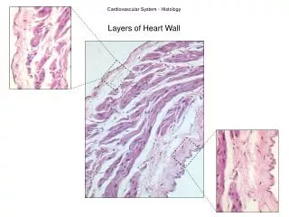
THE WALL OF THE HEART Epicardium = visceral Pericardium (serosa) Myocardium: muscle tissue + c.t. + blood vessels + nerves Endocardium: simple squamous epithelium + c.t. - continuouswith the intima of vessels
EPICARDIUM = visceral pericardium lamina visceralis pericardii serosi · covers the subepicardiac adipose tissue (surrounding the vessels) · flattens the sulci on the surface to prevent friction · reflections PARIETAL PERICARDIUM = fibrous + serous layers • Pericardium fibrosum - collagen + elastic fibrous net , connects to mediastinal pleura,sternopericardiacligg. and the respiratory system • · Pericardium serosum - simple squamous epith, 2 laminae, between them lies the pericardiac cavity (serous surface - frictionless gliding, sliding) • · Lamina visceralis (pericardii serosi) = Epicardium • · Lamina parietalis - lining of the Pericardium fibrosum
MYOCARDIUM Striated, aerobic, interwoven, autorrhythmic Intercalated discs - gap junctions, strong desmosomes Functional syncytium (UNIT = CELL) May undergo hypertrophy Myocardiac infarct - more frequent LEFT SIDE toxicnecrosis - more frequent RIGHT SIDE
LAYERS OF VENTRICULAR MYOCARDIUM EXTERNAL (E) MIDDLE (M) INNER (I) ANULUS FIBROSUS ANULUS FIBROSUS M M M I I E E E I The 3 layered structure is more prominent in the left ventricle
LAYERS OF VENTRICULAR MYOCARDIUM EXTERNAL MIDDLE INTERNAL
ENDOCARDIUM simple squamous epithelium + c.t. - continuouswith the intima of vessels
ENDOCARDIUM Coverspapillarymuscles, trabeculaecarneae chordaetendineae LAYERS Singlelayer of endothelialcells(willtakeup a cuboidalshapeduringsystole) Basementmembrane(loose CT) Subendocardiac CT (connectswiththemyocardium, containselasticnets and branchingsmoothmusclecellstofollowthevolumechanges) Branches of theconductingsystem NO BLOOD VESSELS
BLOOD FLOW PATTERN THROUGH THE HEART • RIGHTATRIUM via the superior and inferior venae cavae • TRICUSPID VALVE into right ventricle • PULMONARY SEMILUNAR VALVES into pulmonary trunk and to the lung • PULMONARY VEINS into the left atrium • BICUSPID VALVE into left ventricle • AORTIC SEMILUNAR VALVES into aorta
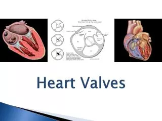
VALVES OF THE HEART 1. 4 SETS OF VALVESPREVENT BACKFLOW OF BLOOD = Mitral valve POSTERIOR P P LEFT RIGHT S A A P B J B J A ANTERIOR Close passively under blood pressure Heart sounds produced by valve closure
ATRIOVENTRICULAR VALVES BICUSPID vs TRICUSPID valves are restrained by chordae tendinae which are in turn attached to papillary muscles (prevention of backflow!)
SEMILUNAR VALVES AORTIC vs PULMONARY AORTA Valvula posterior Valvula sinistra Valvula dextra Lunula Nodulus Pars densa
AUSCULTATION POINTS OF HEART SOUNDS 1st HS: at beginning of ventricular contraction, due to closure of the AV valves 2nd HS: at beginning of ventricular diastole, due to closure of the semilunar valves
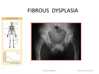
AORTIC STENOSIS PATHOLOGY Poststenotic dilation in the aorta (arrow). Hypertrophy of the left ventricle. NORMAL
MITRAL VALVE PROLAPSE Most common cardiac variation (5-10% of population) Mitral valve cusps do not close properly Regurgitation during left ventricular systole Not life threatening; may be lifestyle threatening