Download
1 / 33
350 likes | 654 Views
Chapter 11 The Muscular System. Skeletal muscle major groupings How movements occur at specific joints Learn the origin, insertion, function and innervation of all major muscles Important to allied health care and physical rehabilitation students. Muscle Attachment Sites: Origin and Insertion.

E N D
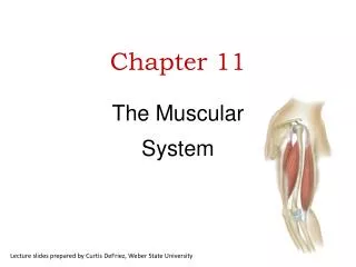
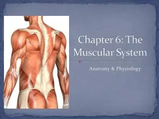
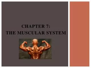
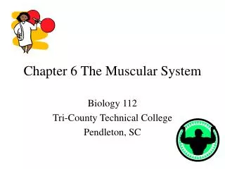
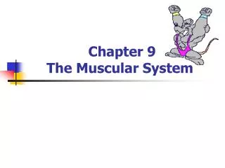
Chapter 11The Muscular System • Skeletal muscle major groupings • How movements occur at specific joints • Learn the origin, insertion, function and innervation of all major muscles • Important to allied health care and physical rehabilitation students
Muscle Attachment Sites:Origin and Insertion • Skeletal muscles shorten & pull on the bones they are attached to • Origin is the bone that does not move when muscle shortens (normally proximal) • Insertion is the movable bone (some 2 joint muscles) • Fleshy portion of the muscle in between attachment sites = belly
Fascicle Arrangements • A contracting muscle shortens to about 70% of its length • Fascicular arrangement represents a compromise between force of contraction (power) and range of motion • muscles with longer fibers have a greater range of motion • a short fiber can contract as forcefully as a long one.
Coordination Within Muscle Groups • Most movement is the result of several muscle working at the same time • Most muscles are arranged in opposing pairs at joints • prime mover or agonist contracts to cause the desired action • antagonist stretches and yields to prime mover • synergists contract to stabilize nearby joints • fixators stabilize the origin of the prime mover • scapula held steady so deltoid can raise arm
How Skeletal Muscle are Named • Direction the muscle fibers run • Size, shape, action, number of origins or locations • Examples triceps brachii -- 3 sites of origin • quadratus femoris -- square shape • serratus anterior -- saw-toothed edge
Muscles of Facial Expression • Arise from skull & insert onto skin • Encircle eyes, nose & mouth • Express emotions • Facial Nerve (VII) • Bell’s palsy = facial paralysis due
Muscles of facial expression • lie within layers of superficial fascia • originate in the fascia or bones of the skull • insert into the skin • move the skin rather than a joint • several act as sphincters • orbicularis oris – closes the eye • levator palpebrae superioris – opens the eye
-sometimes called the occipitofrontalis -dual belly -innervation by VII -two bellies held together by the epicranial aponeurosis Scalp Muscles
Mouth muscles • Orbicularis oris • zygomaticus major & minor • levator labii superioris • depressor labii superioris • depressor anguli oris • levator anguli oris • buccinator • risorius • mentalis
-ZMj: smiling (angle of mouth superiorly and laterally) -ZMr: “snarl” (raises upper lip to expose maxillary teeth)
-risorius: grimace (draws angle laterally)
-DAO: opens the mouth -DLI: depresses lower lip -LLS: raises upper lip -LAO: angle of mouth superiorly & laterally
-buccinator forms the major muscular portion of the cheek -duct of the parotid passes through to reach the oral cavity -compresses the cheeks during blowing
Orbit and Eyebrow Muscles -orbicularis oculi – closes eye -corrugator superioris – frowing -levator palpebrae superioris – opens eye
Extrinsic Muscles of the Eyeballs • Extrinsic muscles insert onto white of eye • Fastest contracting & most precisely controlled • Cranial nerves 3, 4 & 6 innervate the six muscles • 4 Rectus muscles & 2 obliques • Intrinsic muscles are found within the eyeball • strabismus – eyes are unaligned -lazy eye – brain ignores the message sent by one eye and the ignored eye becomes weaker = “amblyopia” -origin of the rectus muscles = common tendinous ring (attached to orbit around the optic foramen) -insert onto the sclera (white) -origin of superior rectus: sphenoid -origin of inferior rectus: maxilla (floor of orbit)
Muscles that Move the Mandible • Masseter, temporalis & pterygoids • Arise from skull & insert on mandible • form part of the TMJ structure • Cranial nerve V (trigeminal nerve) • Protracts, elevates or retracts mandible • Temporalis & Masseter elevate the mandible (biting) • temporalis retracts
-masseter elevates mandible -mandibular division of V -temporalis elevates and retracts mandible -mandibular division of V
Jaw Muscles -- Deep Dissection • Lateral pterygoid protracts mandible • sphenoid bone to condyle of mandible • Medial pterygoid elevates & protracts mandible • sphenoid bone to angle of mandible • Together move jaw side to side to grind food.
Muscles that Move the Tongue • 4 extrinsic muscles ariseelsewhere, but insertinto tongue • Genioglossus • from inside tip of mandible • Styloglossus • from styloid process • Palatoglossus • from hard palate • Hyoglossus • from hyoid bone • Together move tongue in various directions • Intubation is necessary during anesthesia since Genioglossus relaxes & tongue falls posteriorly blocking airway
Muscles of the Floor of the Oral Cavity • Suprahyoid muscles lie superior to hyoid bone. • Digastric m. extends from mandible to mastoid process • used to open the mouth • Mylohyoid m. extends from hyoid to mandible • supports floor of mouth & elevates hyoid bone during swallowing • Stylohyoid & Geniohyoid elevate the hyoid during swallowing
innervated by V and VII – diagastric innervated by V – mylohyoid innervated by VII – stylohyoid innervated by C1 - geniohyoid
Neck muscles • platysma: draws outer part of lower lip inferiorly and pulls skin of chin up as in pouting
Muscles that Move the Head • Sternocleidomastoid muscle • arises from sternum & clavicle & inserts onto mastoid process of skull • innervated by cranial nerve XI (spinal accessory) • contraction of both flexes the cervical vertebrae & extends head • contraction of one, laterally flexes the neck and rotates face in opposite direction
-SCM divides the neck into two triangles 1. anterior triangle: bordered by mandible, sternum, cervical midline & anterior border of SCM -contains the submandibular gland, part of the parotid, the facial artery and vein, common carotids, internal jugular and nerves IX, X, XI and XII -contains three paired sub-triangles + unpaired submental a. submandibular b. carotid c. muscular 2. posterior triangle: bordered by clavicle, SCM and anteriorly by the trapezius -subdivided into: a. occipital b. supraclavicular -contains part of external jugular, subclavian artery, brachial plexus and nerve XI
Muscles that Move the Vertebrae • Quite complex due to overlap • Erector spinae fibers run longitudinally • 3 groupings • spinalis • iliocostalis • longissimus • extend vertebral column • Smaller, deeper muscles • transversospinalis group • semispinalis, multifidis & rotatores • run from transverse process to dorsal spine of vertebrae above & help rotate vertebrae
Scalene Muscle Group • Attach cervical vertebrae to uppermost ribs • Flex, laterally flex & rotate the head