Download
1 / 42
430 likes | 524 Views
The Cardiovascular System: The Heart. Beats approximately 100,000 x/ day Beating 3 billion x/ 70 yr life Over 100,000 km of blood vessels Total blood volume in an average adult is 5L. Functions of the Cardiorespiratory System. Protection Transportation Regulation Gas exchange

E N D
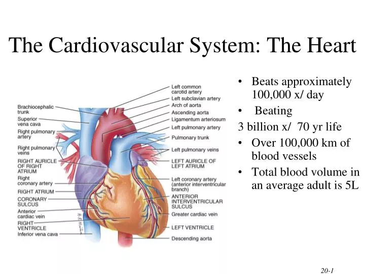
The Cardiovascular System: The Heart • Beats approximately 100,000 x/ day • Beating 3 billion x/ 70 yr life • Over 100,000 km of blood vessels • Total blood volume in an average adult is 5L
Functions of the Cardiorespiratory System • Protection • Transportation • Regulation • Gas exchange • Air purifier
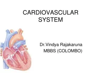
General Characteristics of the Heart • Size of a closed fist • located in thoracic cavity between lungs - mediastiniun • 2 upper chambers - atrium • 2 lower chambers - ventricles • each set separated by a septum • Right side deals with deoxygenated blood • Left side deals with oxygenated blood
The Heart’s Linings • Pericardial sac filled with fluid to reduce friction(dense irregular CT) • Epicardium - outer lining of heart • Myocardium is the heart muscle • Endocardium - lines the inside of the heart
Chambers of the Heart • Two Atria • Right atrium gets deoxygenated blood from the superior and inferior vena cava • Left atrium gets oxygenated blood from pulmonary veins • Two Ventricles • Left has thicker wall and pumps to the body • Right pumps blood to lungs to get oxygenated • Separated by interventricular septum
The Four Valves of the Heart • Atrioventricular valves (gateway to ventricles) • Right = tricuspid; • Left = bicuspid/mitral • Cusps attached to papillary muscles by chordae tendinae • Leaks = murmurs • Semilunar valves • gateway to lungs (puulmonary) • and aorta (aortic)
Valve Function A-V Valves Atria contract, blood fills ventricles through A-V valves SL Valves Ventricles contract, blood pumped into aorta and pulmonary trunk through SL valves
Blood Circulation • Blood flow • blue = deoxygenated (R) • red = oxygenated (L)
Superior and inferior vena cava Right atrium Tricuspid valve Right ventricle Pulmonary valve Pulmonary trunk Gas exchange in lungs Pulmonary veins Left atrium Mitral valve Left ventricle Aortic valve Aorta Gas exchange with working cells Path of Blood through the Heart
Venules and Veins Carries blood towards the heart Usually carries deoxygenated blood except for the pulmonary vein Major properties limited contractibility and elasticity One-way valves (varicose veins) Arteries and Arterioles Carries blood away from theheart Usually carries oxygenated blood except for the pulmonary artery Thick smooth muscle wall Major properties Contractibility Elasticity The Vascular System
The Vascular System Capillaries • Permit exchange of nutrients and gases; walls are one cell thick • Capillaries connect arterioles and venules
Skeletal Muscle Pump Bringing Blood Back to the Heart • Three main ways: • Thoracic pump • Venoconstriction • Skeletal muscle pump • muscle contraction • one-way valves
Coronary Circulation • Right and left coronary arteries nourish the myocardium (heart muscle) • Left and right cardiac veins remove waste from the myocardium
Conduction System of Heart Coordinates contraction of heart muscle
SA node (90-100 x/ minute) cluster of cells in wall of Rt. Atria that fire an electrical pulse begins heart activity that spreads to both atria excitation spreads to AV node AV node (40-50 times x/ minute) in atrial septum (dividing both atria) transmits signal to bundle of His delays the impulse to allow atria to fully contract Bundle of His & Purkinje Fibers the connection between atria and ventricles (via septum) divides into bundle branches & purkinje fibers, large diameter fibers that conduct signals quickly Electrical Conduction of Heart
Rhythm of Conduction System • SA node fires spontaneously 90-100 times per minute • SA node setting pace since is the fastest • AV node fires at 40-50 times per minute • If both nodes are suppressed fibers in ventricles by themselves fire only 20-40 times per minute • Artificial pacemaker needed if pace is too slow • Note: • caffeine & nicotine increase activity
Electrocardiogram---ECG or EKG • EKG • Action potentials of all active cells can be detected and recorded • P wave =Atrial Depolarization • spreads from the SA nodethrough the atria • 0.1s after the P wave begins, atria contracts • repolarization of atria not evident because it is buried in the QRS complex • P to Q interval • conduction time from atrial to ventricular excitation
Electrocardiogram---ECG or EKG • QRS complex = Ventricular Depolarization - shortly after QRS wave begins, the ventricles contract • T wave = Ventricular Repolarization • ventricular repolarization • occurs before the ventricles start to relax • smaller & more spread out because repolarization takes longer
Abnormal ECG/ EKG • Large P Wave = Enlarged Atria - problems with the bi or tricuspid valves causes a backup of blood in the atria resulting in the expansion of the atrial walls • Enlarged Q Wave = Myocardial Infarction (HEART ATACK!!) • Enlarged R wave = Enlarged Ventricles • Flatter T Wave = The Heart receiving insufficient Oxygen • Tachycardia = a fast resting heart beat greater than 100bpm in adults • Bradycardia=an abnormally slow/unsteady resting heart rate < 50bpm
Heart Sounds Where to listen on chest wall for heart sounds.
Systole and Diastole • Cardiac cycle • Systole when ventricles contract (heart empties) • Diastole when ventricles relax (heart fills) • Heart sounds heard through a stethoscope Lub - a “long/low” sound - closing of the a-v valves (tri/bi) Dub - A “sort/sharp” sound closing of the s-v (aorta/ pulmonary)
Cardiac Output • Amount of blood pushed into aorta or pulmonary trunk by ventricle • Determined by stroke volume and heart rate • CO = SV x HR • at 70ml stroke volume & 75 beat/min----5.25 L/min • entire blood supply passes through circulatory system every minute • Cardiac reserve is maximum output/output at rest • average is 4-5 L/ min while athlete is 7-8 L/ min
Cardiac Output Calulations Example: HR = 70 bpm SV = 70 mL (Q) CO = HR x SV = 70 beats/ min x 70 mL of blood/ beat = 5040 mL/ min = 5.04 L/ min
Factors Affecting Heart Rate • Age – child’s HR much faster than adult • Emotional State of the Individual • Parasympathetic Nervous System HR • Sympathetic Nervous System HR 3) The physical state & efficiency of the heart • Athletic heart has larger SV & lower RHR • Couch Potato has a faster RHR
Regulation of Heart Rate • Nervous control from the cardiovascular center in the medulla • Sympathetic impulses ↑ heart rate & contraction • parasympathetic impulses ↓ heart rate. • Baroreceptors (pressure receptors) detect change in BP and send info to the cardiovascular center
Regulation of Heart Rate • Heart rate is also affected by hormones • epinephrine, norepinephrine, thyroid hormones • ions (Na+, K+, Ca2+) • age, gender, physical fitness, and temperature
Influences on Stroke Volume • Preload (affect of stretching) • Frank-Starling Law of Heart • more muscle is stretched,greater contraction force • more blood more force of contraction results • Contractility • autonomic nerves (stress), hormones a) Contractility = Parasympathetic Stimulation • b) Contractility = Sympathetic Stimulation • Afterload • amount of pressure created by the blood in the way • high blood pressure creates high afterload
CV Systems’ Adaptation to Exercise • With improved CV fitness • SV will increase (increased mass & contractibility) • therefore, RHR will decrease • Also the Max CO will increase (VO2 Max will also increase due to this) • RBP will become more constant (120/80) • Rick of CV diseases will decrease • Increased # of capillaries around the heart
CV Systems’ Adaptation to Exercise • Increased Myoglobin (02 binding pigment) • acts as an 02 store aiding in the diffusion of 02 2) Increased oxidation of carbohydrates • training increases the muscles capacity to break down glycogen in the presence of 02 3) Increased oxidation of fats - training increases the muscles capacity to break down fatty acids in the presence of 02
VO2 Max Definition: the max amount of O2 that can be consumed per minute during max exercise (measured in mL/ kg) • also known as aerobic power • this is an individuals max aerobic capacity, or ability to consume O2 at the cellular level
VO2 Max • 93% of VO2 Max is under genetic influence, although it can be improved through training, there is a genetic ceiling • Max VO2 doesn’t differ between boys & girls before puberty, after puberty females are 25 – 230% less than values
VO2 Max • Capacity depends on the amount of O2 that can be delivered to the muscles compared to the amount of O2 used by the muscle • O2 consumption is important to prolonged exercise. (Endurance activities such as marathons, triathletes)
Blood Pressure Blood pressure refers to the force exerted by circulating blood on the walls of blood vessels • Systolic - The force your blood exerts when the heart is contracting • Diastolic - The force your blood exerts when the heat is relaxing • Measured using a sphygmanometer
Blood Pressure • Factors affecting blood pressure • Cardiac output • Peripheral resistance • Blood volume • Blood pressure • Normal = 120/80mmHg • Hypertensive = 140/90mmHg
Hypertension -persistently elevated blood pressure - a major cause of heart failure, kidney failure, & stroke Risk Factors: • 1)Sex – male • 2)Race – Black • 3)Lifestyle – smoker, diet, drinker 4) Genetics – hypercholesterolemia, glucose intolerance (diabetic)
Myocardial Infarctions/ Attacks • Blockage of a coronary artery (due to plaque & fat) depriving the heart of O2 • Tissue in the affected area suffers permanent injury & signals its distress by a very sharp pain (angina) • If the damage to the heart muscle is too extensive, the individual will not survive
Myocardial Infarctions/ Attacks • Signs: pressure in the chest that lasts more than a few minutes, or goes away comes back • Spreading pain – shoulders, neck, left arm • Lightheaded, sweating, nausea
Angina Pectoris • Chest pain due to CHD • Ischemia (insufficient blood supply) • Occurs when blood flow to the heart doesn’t meet increased demands Rx: take nitroglyerin • relaxes the veins ( amount of venous return, work of the heart) • relaxes the coronary arteries ( the amount of blood supplied to the heart)