Download
1 / 56
750 likes | 1.31k Views
Peritoneal Dialysis. Kampol Pattanasombatsakul. Peritoneal Anatomy. Peritoneum: 1-1.3 m 2 Visceral: 90% Parietal: 10% Peritoneal cavity Potential space 10-100 ml Vascular supply. Peritoneal Physiology. Peritoneal Dialysis จะเริ่มอย่างไร. Prepare unit for PD patients.

E N D
Peritoneal Dialysis KampolPattanasombatsakul
Peritoneal Anatomy • Peritoneum: 1-1.3 m2 • Visceral: 90% • Parietal:10% • Peritoneal cavity • Potential space • 10-100 ml • Vascular supply
Peritoneal Dialysisจะเริ่มอย่างไร
Prepare unit for PD patients • Personal (Team) • Nephrologist , Surgeon • Nurse , on call nurse • Other • Equipment • In unit : washing basin, dressing set, catheter , connector , dialysis set , • At home : washing basin, storage , clean towel , weight • Management • Education tools • Data management , Statistic • First visit , hospitalization ,
Home Care therapy : benefit to Persons involved in dialysis care team
Prepare unit for PD patients • Personal • Nephrologist , Surgeon • Nurse , on call nurse • Other • Equipment • In unit : washing basin, dressing set, catheter , connector , dialysis set , • At home : washing basin, storage , clean towel , weight • Management • Education tools • Data management , Statistic • First visit , hospitalization ,
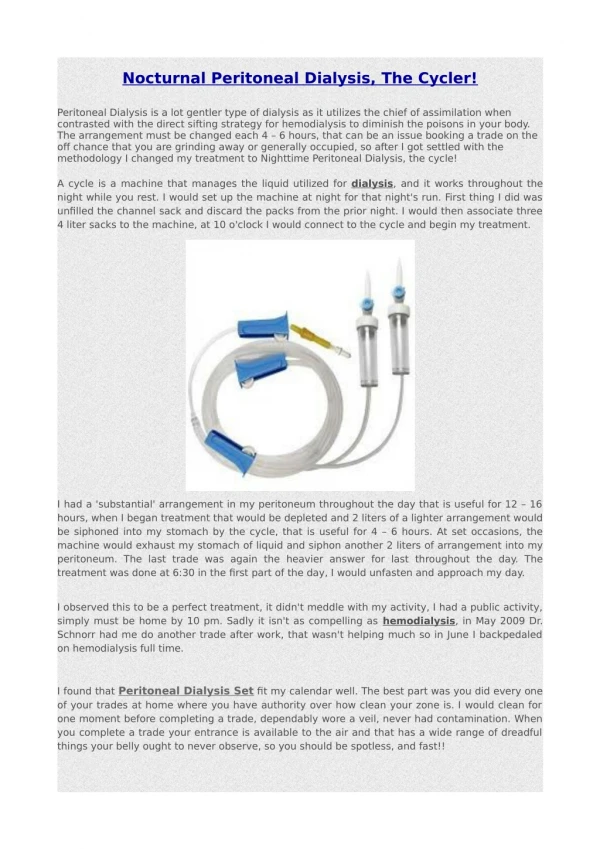
Different PD catheters Straight Tenckhoff catheters Coiled Tenckhoff catheters Straight 1 cuff Straight 2 cuffs Coiled 1 cuff Coiled 2 cuffs Swan Neck Tenckhoff catheters Swan Neck Missouri Coiled Coiled Straight Straight Bead placed IP, Flange extraP • Downwards directed exit site • Permanent bend between 2 cuffs (180°) • Right or left
Prepare unit for PD patients • Personal • Nephrologist , Surgeon • Nurse , on call nurse • Other • Equipment • In unit : washing basin, dressing set, catheter , connector , dialysis set , • At home : washing basin, storage , clean towel , weight • Management • Education tools • Data management , Statistic • First visit , hospitalization ,
Education tool • CKD, kidney function • Catheter care and aseptic technique • Dialysis exchange • Fluid and nutritional balance • Record • Complication • Medication • Follow up
Select the patients • Not suitable for PD • Skin disease over abdomen • Serious abdominal surgery • Malnourish : S. albumin • Large BSA > 2.0 • Very low GFR < 2 ml/min • Abdominal anatomical defect • ? COPD • ? Diabetic
Theoretically not to choose PD initially –BUT PD may be feasible with added adjustments • Large body size • Diverticulosis / diverticulitis • Severe backache NIPD • Hernias NIPD • Multiple abdominal surgery • Poor manual dexterity • Blindness • No compliance
PD preferred • Bleeding diathesis (no need of heparinization) • Diabetes (status of vessels, insulin i.p.) • Chronic infections (prevention of the nosocomial spread – hepatitis B, C, HIV) • Future transplantation (improved initial graft function rate) • Multiple myeloma (improves the chances of renal recovery, removes some light-chains proteins)
PD and HD equally preferred • Polycystic kidney disease • Scleroderma, other conective tissue diseases (e.g. SLE) • Patients living in nursing homes
PD preferred Independent Life Frequent travels Tendency towards PD Great need of independence by the patient Need to maintain work Distance to the HD center Psychosocial situations in which PD is more appropriate
Prepare the patients • Who choose method of renal replacement therapy ? • Who will take care the patients? • Uremic symptoms • Volume balance • Compliance • Socioeconomic and hygiene status
CAPD for a new patient • Patient education • Patient training program • Catheter placement • Break – in period & training time • Peritoneal testing • Mode of Dialysis
Patient Education ๑.ไตวายเรื้อรัง และ ไตวายระยะสุดท้าย ๒. การควบคุมอาหาร น้ำ และ การรับประทานยา ๓. อาการและอาการแสดงต่างๆ ๔. ภาวะแทรกซ้อน และ ความผิดปกติที่พบได้ ๕. ความสำคัญของการบำบัดไตทดแทน
Patient training program • Hand wash • Basic sterile technique • Exchange procedure • การทำแผล ( exit site care ) • การฉีดยาเข้าถุงน้ำยา • การเก็บน้ำยาส่งตรวจ
Peritoneal Dialysis-Related InfectionsRecommendations : 2005 update Catheter placement • Pre operative preparation • Ideal location should be mark by team • Free of constipation • Controversy : nose culture for S. aureus nasal carrier, if positive , 5-day course intranasal mupirocin will be used. • Operative • Prophylaxis ABO (evidence): 1st generation cephalosporin ( randomized control : vancomycin 1 gm iv, single dose is superior than cephalosporin ) ( odd ratio 11.5 : 6.45 : 1) • Using Double cuff with superficial cuff should be 2-3 cm. from the exit site • Downward direct tunnel • Swan neck catheter ( not confirm ) • Avoid trauma and hematoma during catheter placement • Suture increase risk of infection and are contraindicate PDI 2005: 25(2) ; 107-131
Peritoneal catheter Exit site Cuffs
Post operative care • Pain management • Antibiotic • Break in period : rinse peritoneum • ? Dialysis need • Consider HD first • Hospitalization • Training review , catheter exchange system
Immediate post operative state • Perioperative pain • Bloody effluent • Dialysate leakage • Displacement • Perioperative peritonitis • Temporary obstruction • Cuff extrusion
Immediate post operative state Perioperative pain • Wound pain • Catheter related pain – reaction , location • In flow pain– dialysate related • Management • Adequate analgesic during operation and post operative period • Pseudo ascites during catheter insertion • No guide-wire / smooth catheter insertion • Slow infusion / low volume • Non osmotic dialysate solution • Warm dialysate
Immediate post operative state Bloody Effluent • Bleeding complication during surgical procedure • Bleeding tendency in end stage renal disease • Thrombocytopenia • Management • Rinse peritoneal cavity until clear • Add heparin 500-1000 unit / L in last bag dialysate • Correction of bleeding tendency • Peritoneal resting • Prevention
Immediate post operative state Dialysate leakage • Leakage via insertion point, exit site • Commonly found in midline insertion, acute PD catheter • How to test – only dipstick for glucose • Management • Prevention – catheter fixation first 2-3 days , soak both cuffs with NSS • Decrease intraabdominal pressure – using low volume , avoid constipation • Temporary hemodialysis • Late catheter use
Immediate post operative state Displacement • Upward displacement of catheter , non in appropiated position • How to diagnose – plain abdomen • Management • Prevention – constipation • During catheter insertion – pain management , deep cuff fixation , subcutaneous tract • Laxtive along with early ambulation • Guide wire manipulation • Re-exploration
Immediate post operative state Perioperative peritonitis • Early peritonitis • Cause – infection , reaction( plasticizer, air , fluid component ) • How to diagnose – cell count & differential ( PMN , Eosinophil ) • Management • Prevention – antibiotic prophylaxis • Sterile technique • Wound dressing • Antibiotic , prefere i.v. for infection • Symptomatic treatment for reaction / allergy – usually resolve in 2 weeks
Immediate post operative state Temporary obstruction • Inflow obstruction – fibrin , clot • Outflow obstruction ( one way valve ) – omentum occlusion , catheter clinking, displacement • Complete obstruction • Management • Prevention –gently catheter insertion without guide wire , control bleeding before insertion , catheter testing immediate insertion , pseudoascites • NSS flush , intraluminal fibrinolytic agents, heparin locked catheter, brush , guidewire manipulation • NSS 100 ml + heparin 1000 u intraperitoneally , peritoneum resting • Guidewire manipulation , reinsertion
Before starting PD • Make sure dialysis exchange and septic technique • Select type of dialysis solution • Do we need PET? • Select mode of dialysis • Select dialysis dose
Volume : Guideline • Acute dialysis • small volume : 1000 ml • increase 1500 -2000 ml in supine position • Chronic dialysis • volume 2000 - 3000 ml
Peritoneal dialysis solution • Buffer compositions • Lactate 35 - 40 mEq/L • Bicarbonate 25 - 39 mEq/L • Osmotic compositions • Glucose ( Dextrose ) : 1.36% , 2.25% , 3.86% glucose 1.5% , 2.5% , 4.25% dextrose • Electrolytes • Sodium : 132 - 134 mEq/L dialysate • Magnesium : 0.5 -1.0 mEq/L • Potassium : 0 - 2.0 mEq/L • Calcium : 2.0 - 3.5 mEq/L ( 1.0 - 1.75 mmol/ L)
PD solution : Calcium • 2.0 - 3.5 mEq/L ( 1.0 - 1.75 mmol/L ) • Precaution of low calcium dialysate • Negative calcium balance • induce severe hyperparathyroidism • Indication for low calcium dialysate ( 2.0 mEq/L ) • hypercalcemia • serum phosphate > 3.0 mEq/L with oral calcium • serum phosphate < 3.0 mEq/L with hyperparathyroidism with oral vitamin D • hypoparathyroidism
Ectopic Calcification • despite better P control, PD patients may be at increased risk for vascular calcification because of low turnover bone • vascular medial calcification associated with arterial stiffening and increased afterload • vascular intimal calcification associated with luminal narrowing and occlusive disease
Fate of Ingested Calcium – Normal or Increased Bone Turnover Bone uptake Oral Ca Gut absorption Visceral Calc’n Vascular Calc’n
Fate of Ingested Calcium – Low Bone Turnover Bone uptake Oral Ca Gut absorption Visceral Calc’n Vascular Calc’n
Medial Calcification Intimal Calcification • produces stiff arteries • may be responsible for LVH and CHF • tends to correlate with plaque area • may be responsible for occlusive vascular disease
How to diagnosed adynamic bone disease in dialysis patients? • Bone marker • Bone turnover Parathyroid hormone(<150 pg.ml) • Resorption Tartarate resistant acid phosphatase ( TRAP) Pyridinoline and deoxypyridinoline ( PYD,DPD) Procollagen type I cross linked C-terminal telopeptide ( ICTP) • Formation : Total alkaline phosphatase ( TAP) Bone alkaline phosphatase ( BAP) < 27 U/L Osteoclacin ( OC) < 14 ng/ml Procollagen type I carboxy terminal propeptide ( PICP) • Bone biopsy
Before starting PD • Make sure dialysis exchange and septic technique • Select type of dialysis solution • Do we need PET? • Select mode of dialysis • Select dialysis dose
Standard PET • Upright , drain the overnight (8-12hr) over 20 min. • Supine , infuse 2.0 L , 2.5 %D in 10 min. Roll side by side every 400 ml or 2 min. • Complete ( Zero time ) and 2hr dwell , drain 200 ml , mix well , collect 10 ml of dialysate , reinfuse 190 ml • Collect blood at 2 hr. • At 4 hr. drain all fluid , mix well , measure volume and collect sample
Peritoneal Equilibration Test Solute clearance Ultrafiltration
Before starting PD • Make sure dialysis exchange and septic technique • Select type of dialysis solution • Do we need PET? • Select mode of dialysis • Select dialysis dose
CAPD prescription • CAPD (continuous ambulatory PD) • NIPD ( nocturnal intermittent PD) • CCPD ( continuous cyclical PD) • DAPD ( day time ambulatory PD)
Adequacy recommendation Method Weekly Kt/V Creatinine clearance ( L/wk/1.73sq.m.) CAPD >2.0 >60 NIPD >66 >2.2 >63 CCPD >2.1 • Calculation of renal creatinine clearance for 24 hours: • Total urinary creatinine removal divided by serum creatinine level • U/Pcreatinine x urine volume • Standardized by body surface area • Calculation of peritoneal clearance (Urea/Creatinine) for 24 hours • Total peritoneal removal divided by serum level • D/P x total dialysate volume • Standardized by body surface area (creatinine)-Peritoneal creatinine clearance • Standardized by body water content (urea): Kt/Vurea
Adequacy recommendation At least Kt/V > 1.7 in CAPD ; At least Kt/V 1.7 & CCr 45 L/wk/1.73 sq.m for APD Krt/V 2.0 = GFR ประมาณ 9.38 – 12.43 ml/min Estimate GFR = - 0.4462 + ( 4.9179 * Krt/V ); หรือ Krt/V= 0.09 + estimate GFR /4.9179
Before starting PD • Make sure dialysis exchange and septic technique • Select type of dialysis solution • Do we need PET? • Select mode of dialysis • Select dialysis dose
PD prescription Determine RRF Low RRF, urine vol High RRF Start with NIPD, may be with 3 x 2L Start with CAPD, 4 x 2L unless very small size Inadequate UF/clearance Inadequate Clearance Inadequate UF High transport Low transport Inadequate clearance Yet adequate UF High transport高转运 Increase dwell vol/ mid-day ex.CCPD ?? PD+HD CCPD/NIPD + mid-day exchange NIPD with 4 x 2 L, Or 5 x 2L Increase volume / frequency, day dwell