Download
1 / 8
80 likes | 220 Views
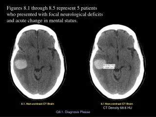
Contrast-enhanced Dedicated Breast CT: Initial Clinical Experience. Radiology. 2010 September; 256(3): 714 – 723. Purpose.

E N D
Contrast-enhanced Dedicated Breast CT: Initial Clinical Experience Radiology. 2010 September; 256(3): 714–723
Purpose • To quantify contrast material enhancement of breast lesions scanned with dedicated breast computed tomography (CT) and to compare their conspicuity with that at unenhanced breast CT and mammography
Materials andMethods • 46 women (mean age, 53.2 years; age range, 35–72 years) with Breast Imaging Reporting and Data System category 4 or 5 lesions underwent unenhanced breast CT and contrast material–enhanced breast CT before biopsy. Two radiologists independently scored lesion conspicuity for contrast-enhanced breast CT versus mammography and for contrast-enhanced breast CT versus unenhanced breast CT. Mean lesion voxel intensity was measured in Hounsfield units and normalized to adipose tissue intensity on manually segmented images obtained before and after administration of contrast material. Regression models focused on conspicuity and quantified enhancement were used to estimate the effect of pathologic diagnosis (benign vs malignant), lesion type (mass vs calcifications), breast density, and interradiologist variability
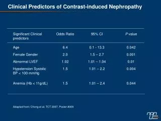
Results • Fifty-four lesions (25 benign, 29 malignant) in 46 subjects were analyzed. Malignant lesions were seen significantly better at contrast-enhanced breast CT than at unenhanced breast CT ( P <0.001) or mammography ( P <0.001). Malignant calcifications (malignant lesions manifested mammographically as microcalcifications only, n = 7) were seen better at contrast-enhanced breast CT than at unenhanced breast CT ( P <.001) and were seen similarly at contrast-enhanced breast CT and mammography. Malignant lesions enhanced 55.9 HU +_ 4.0 (standard error), whereas benign lesions enhanced 17.6 HU+_ 6.1 ( P < .001). Ductal carcinoma in situ ( n = 5) (DCIS) enhanced a mean of 59.6 HU+- 2.8.
Conclusion • Conspicuity of malignant breast lesions, including ductal carcinoma in situ, is significantly improved at contrast-enhanced breast CT. Quantifying lesion enhancement may aid in the detection and diagnosis of breast cancer.
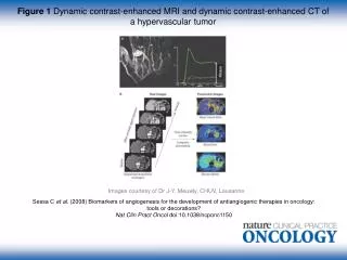
Images obtained in a 48-year-old woman. (a) Implant-displacedcraniocaudal mammogram shows a mostly obscured mass. (b)Pre- and (c) postcontrast coronal breast CT images show the same invasive mammary carcinoma mass that enhanced 76.5 HU
(b) precontrast coronal, (c) post-contrast coronal,(d) post-contrast sagittal, and (e) Post-contrast transverse breast CT images show the same focus of DCIS that enhanced 50.2 HU (a)Mediolateral oblique magnification mamm-ogram shows a group of indeterminate microcalcifications.
(a) Postcontrast transverse, (b) precontrast coronal, and (c) postcontrast coronal breast CT images of a mucinous adenocarcinoma (arrowheads) with partial rim enhancement (arrow) in a 46-year-old woman.