Download
1 / 46
460 likes | 468 Views
Perbanyakan Massal Virus sebagai Agens Antagonis. By Irda Safni. Virus sebagai agens antagonis patogen tumbuhan. Ada beberapa cara virus dapat digunakan untuk mengendalikan patogen tumbuhan, yaitu :

E N D
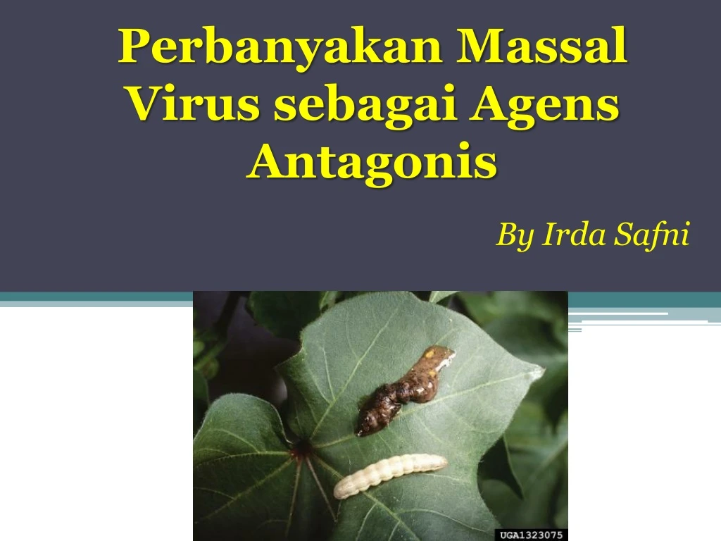
Perbanyakan Massal Virus sebagai Agens Antagonis By Irda Safni
Ada beberapa cara virus dapat digunakan untuk mengendalikan patogen tumbuhan, yaitu : 1. Virus dapat membunuh atau mengurangi patogenisitas bakteri patogen dan jamur patogen tumbuhan. 2. Virus dapat membunuh vektor invertebrata virus pada tanaman tingkat tinggi sehingga mencegah penyebarannya. 3. Strain yang agak lemah yang menyebabkan sedikit atau tidak menimbulkan dapat melindungi tanaman dari infeksi laten oleh strain yang lebih parah.
Stanway (1985) mendeteksi infeksi virus infection sebanyak 126 dari 157 isolat jamur take-all pada tanaman gandum (Gaeumannomyces graminis var. Tritici). • Kultivar gandum yang terinfeksi Helminthosporium victoriae (penyakit hawar) adalah penyakit penting gandum di Amerikapada 1947 and 1948. • Lindberg(1959) menemukan beberapa koloni jamur H. victoriae menjadi memendek, ditandai dengan daerah pada pinggir koloni, miselium udara menjadi lisis dan menghambat perkembangan koloni. • Penyakit ini juga ditransmisikan ke kultur yang sehat oleh anastomosis hifa dan mungkin disebabkan oleh 1 atau 2 dsRNA virus yang biasa dijumpai padaH. victoriae (Ghabrial 1986). • Tanaman gandum yang dijumpai isolat jamur tidak menyebabkan kehilangan hasil yang besar, kemungkinan disebabkan penyebaran virus pada populasi H. Victoriae, karena isolat menghasilkan sedikit toksin victorin dan menjadi kurang patogenik dibanding isolat yang normal (Lindberg 1960).
Isolat kultur Rhizoctonia solani mengandung 3 segmen virus dsRNA, sedangkan tidak dijumpai virus dsRNA pada ujung hifa yang sehat dari isolat yang mengifeksi.
Penyakit yang disebabkan virus entomopatogen mulai diketahui sejak abad ke-16. • Penyakit yang disebut Jaundice o graserrie, sekarang diidentifikasi sebagai nucleopolyhedrosis, ditemukan pada ulat sutra (Bobyx mory) oleh Vida pada tahun 1524 dan kemudian juga diisolasi dari lebah madu (Apis mellifera). • Pada tahun 1856, dua orang ahli Italia (Maestri dan Cornalia) menjelaskan occlusion bodies (OBs) ulat sutra nucleopolyhedrosis. • Pada tahun 1926 Paillot mendeskripsikan granulovirus (GVs) pertama sekali. • Pada tahun 1934 Ishimori menjelaskan jenis baru polyhedrosis di dalam ulat sutra OBs dibentuk didalam sitoplasma sel yang diinfeksi (bukan pada asam nukleat) sekarang dikenal dengan cypovirus.
Sejak tahun 1950 s/d 1970, Steinhaus dan koleganya menguji baculovirus sebagai agens hayati di lapangan dengan mengaplikasi nucleopolyhedrovirus (NPV) untuk mengendalikan ulat alfalfa (Colias eurytheme Boisduval; Lepidoptera: Pieridae). • Bioinsektisida komersil berbahan aktif virus pertama dikembangkan pertama sekali pada tahun 1975 oleh Perusahaan Sandoz (dengan nama dagang Elcar) untuk mengendalikan Heliothis/Helicoverpa Lepidoptera: Noctuidae). • Selama tahun 1979 s/d 1980, penemuan penting pada genetika virus entomopatogen, khususnya baculovirus. • Hingga saat ini studi genetika virus entomopatogen difokuskan pada studi genom lengkap telah ada 29 sekuensing genom lengkap virus entomopatogen.
Keuntungan dan Kerugian Penggunaan Virus untuk mengendalikan hama Keuntungan Selektif dan efektif terhadap hama sasaran. Aman bagi serangga dan organisme bukan sasaran serta tidak menyebabkan resistensi. Persisten dan tidak meninggalkan residu beracun di alam. Tidak menyebabkan pencemaran lingkungan. Efektif menginfeksi ulat yang telah terkena insektisida kimia. Tidak menyebabkan peningkatan populasi hama sekunder. Dapat ditularkan oleh parasitoid dan predator ke inang yang sehat. Dapat mengendalikan ulat instar V-VI. Tidak menyebabkan penyakit virus pada tanaman. Kompatibel dengan teknik pengendalian yang lain, termasuk insektisida kimia. Mudah diproduksi dengan teknik sederhana (menggunakan alat semprot standar). Berpotensi sebagai pengendali hama jangka panjang. Dapat beradaptasi dengan teknologi modifikasi secara genetik (bioteknologi).
Kerugian • Hanya spesifik terhadap hama sasaran. • Kemungkinan berbahaya bagi serangga bukan sasaran. • Waktu aplikasi harus tepat untuk memaksimalkan efektivitas. • Memerlukan pendistribusian secara merata pada kanopi tanaman untuk meningkatkan kontak dengan hama sasaran. • Daya bunuh lambat. • Rentan terhadap pengaruh lingkungan. • Kehilangan virulensi dan patogenitas jika diperbanyak secara terus menerus (jika tidak dilakukan penggantian inang baru). • Infektivitasnya di lapangan singkat dan membutuhkan penanganan tertentu. • Kekhawatiran masyarakat terhadap kemungkinan patogenik/menyebabkan alergi. • Menurunkan dosis perlakuan melalui bioteknologi menyebabkan resurgensi hama atau serangga bukan sasaran.
Saran untuk aplikasi virus entomopatogen • Virus tidak dapat diaplikasikan sendiri, tetapi kerkonjugasi dengan teknik pengendalian yang lain. • Virus entomopatogen bersifat spesifik, sehingga serangga target farus diidentifikasi secara benar. • Lahan dicangkul terlebih dahulu sebelum aplikasi dilakukan, dan virus diaplikasikan pada serangga target sewaktu masih mudah tetapi aktif makan.
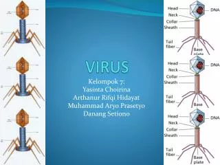
Virus yang Menginfeksi Invertebrata • Virus DNA • Double stranded DNA • - Poxviridae • - Iridoviridae • - Baculoviridae: NPV & GV • - Polydnaviridae • Single stranded DNA • - Parvoviridae • Virus RNA • Double stranded RNA • - Rheoviridae: Cypovirus • Single stranded RNA (-) • - Rhabdoviridae • - Bunyaviridae • Single stranded RNA (+) • - Picornaviridae, Togaviridae, • Tetraviridae, Flaviridiae, • Nodaviridae
Tabel 1. Kelompok Virus Entomopatogen ds= double-stranded, ss=single-stranded.
6 Baculoviruses Spodopteralittoralis 2microns FromHunter-Fujitaetal
7 Baculoviruses- Modeofaction FromHunter-Fujitaetal
8 Baculoviruses SusceptibilityofAlternativeHosts Foundonlyininvertebrates Nomemberofthefamilyisknowntoinfectplantor vertebrate Mosthavenarrowhostinsectrange,andinfectivityis restrictedtotheoriginalhostgenusorfamily
9 Baculoviruses Toxicitystudies-mammals Toxicitytestresultsfrom1970s/80sof29NPVs indicatednotoxicityorpathogenicity.Doseswere generally10–100xthe“peracre”(1acre=0.45ha) fieldrateequatedtoa70kgperson. HeliothiszeaNPVmostextensivelytestedfortoxicityin humansandledtoregistrationof“Elcar”bySandozin USA.
10 Baculoviruses Toxicitystudies–mammalscont. NoeffectsofHzNPVfoundin: Acutetoxicity-pathogenicitytestsinmouse,rat,guineapig,rabbit, monkeyandmanat6x109–3x10¹²OB/kg. Skinirritationsensitivitytestsinguineapigs,rabbitsandmanat106 and107OB/mm2skin. Eyeirritationtestsinrabbitswith105and2x106OB/eye Subacutetoxicity-pathogenicitytestsandsubcutaneousinjectioninto mice,rats,dogsandrhesusmonkeys. Teratogenicityandcarcenogenicitystudiesinratsandmiceat109– 3.5x10¹²OB/kg. SimilarbutlessextensiveresultsformanyotherNPVsfromthe 1970s/80s
11 Baculoviruses Toxicitystudies–wildlife Birds AbletopassNPVthroughthealimentarytractunaffected Nodeleteriouseffects Aquaticorganisms Noadverseeffects Beneficialinsects Nodirecteffectonparasitoids,predatorsandpollinators Indirecteffectsonparasitoidsresultingfromhostdeath
12 Baculoviruses Pathologystudies Toxicitytestsdesignedfortestingeffectsofchemicalson vertebratesareinsufficient ResultsreportedinGröner(1986)indicatenovirusinduced antibodyproductionintestmammalsandchicken. Nocytogeneticeffectsofbaculovirusesinmammaliancellseither invivoorinvitro.
13 Baculoviruses Virus-cellinteractionsinvitro AcNPVinoculatedintovertebratecellscanbetaken- upandthedegreeofup-takedependsoncelltype, temperature,timeandviralphenotype. BUT,noneofthehumanandnonhumanvertebrate linestestedshowedevidenceofviralreplication. NPVsunabletoactivateretrovirusesinmammalian celllines
15 Baculoviruses alistofthebaculovirusesregulatedaspesticide activeingredientsbytheUSEPAOfficeofPesticide ProgramsasofMay2005 AnagraphafalciferaNPV CydiapomonellaGV DouglasfirtussockmothNPV GypsymothNPV HelicoverpazeaNPV IndianmealmothGV MamestraconfigurataNPV(pending) SpodopteraexiguaNPV
16 Baculoviruses–USEPAfactsheet III.ASSESSINGRISKSTOHUMANHEALTH Thesevirusesinfectonlythetargetinsectlarvaeandcloselyrelated species.Toxicitytestsshowthatthevirusesposenorisktothepublic. Workerswearprotectiveclothingtopreventpossibleirritationfrom handlingandapplyingtheproduct. IV.ASSESSINGRISKSTOTHEENVIRONMENT TestsshowthattheGVandNPVsthatEPAhasregisteredaspesticide activeingredientsspecificallyinfectonlycertainspeciesofmothlarvae. Thevirusesdonotharmotherorganisms,includingplants,beneficial insects,otherwildlife,ortheenvironment.Thesevirusesoccurnaturally intheirinsecthosts.
17 Cypoviruses:Modeofaction Polyhedraingestedanddissolvedinlarvalmidgut Virionsreleasedandattachtomidgutcolumnarcells Viralcoreenterscellcytoplasm RNAtranscriptionandreplication RNAoccludedincapsules Viruscapsulesoccludedbyvirogenicstromatoformocclusion bodies
18 Cypoviruses(Rheoviridae) NoCPVhasbeenfoundinfectingvertebratesorplants (Belloncik,1989) DendrolimusspectabilisCPVregisteredinJapanin 1974.Safetytestresultsgenerallynegative. –Katagiri,K.(1981)Pestcontrolbycytoplasmicpolyhedrososviruses.In: Microbialcontrolofpestsandplantdiseases1970-1980.(EdBurges,H.D.) AcademicPress.
Poxviridae:Entomopoxvirus • Member of the family of Poxviridae has a wide host, including vertebrates and invertebrates. • Chicken pox and Small pox virus belong to this family. • The show allantoid – to brick-shaped virions, occluded within ovoid OBs called Spheroids. • Entomopoxvirus has been isolated from 27 orthopterans, lepidopterans, dipterans and coleopterans. • The subfamily Poxvirinae includes three genera, i.e. EntomopoxvirusA, EntomopoxvirusB, and EntomopoxvirusC • Entomopoxvirus A infects only coleopteran species; EntomopoxvirusB infects lepidopteran and coleopteran species; Entomopoxvirus C infects only dipteran species. • The fourth group, group D, has been proposed by ICTV which only attacks hymenopterans.
Poxviridae:Entomopoxvirus Anomala cuprea (Coleoptera) larvae infected with an entomopoxvirus show the symptoms of the infection such as a whitish appearance and underdevelopment (left, infected larva; right, healthy one).
Ascoviridae: Ascovirus • Members of the family of Ascoviridae are doublestrandeDNA (dsDNA) viruses that infect lepidopteraninsects and cause the unique pathology of forming virioncontainingvesicles in the hemolymph of infected hosts. • The presence of the vesicles gives the hemolymph • a milky white appearance, which is a major characteristic • of the disease. • A few species of Ascovirus has been isolated only from insects, specifically from Lepidopterans (Noctuidae). • Enveloped virions of ascoviruses are bacilliform, ovoid or allantoid in shape, and occluded within vesicle-like OBs.
Ascoviridae: Ascovirus • Ascovirus symptoms • In cases where a Microplitis wasp has both parasitised a caterpillar and infected it with ascovirus, the symptoms seen are those of the disease rather than of the parasitoid.When ascovirus kills the caterpillar, it also kills the developing Microplitis larva. • Caterpillars infected with ascovirus will generally stop eating within two days. They stop growing, but can live for weeks in a lethargic state before they die. • The blood of an ascovirus-infected caterpillar is white and creamy, whereas the blood of a healthy caterpillar is clear. Blood colour gives the best diagnosis in the laboratory and can be tested by splitting or pricking the caterpillar.
Iridoviridae: Iridovirus • Invertebrate Iridescent Viruses (IIVs) (family Iridoviridae) • are known to infect a number of agricultural pests, • medically important insect vectors, and terrestrial isopodsthat live in damp or aquatic habitats. • The major characteristic of this family is the presence of iridescent blue,green, orange, or purple coloration in heavily infected individuals. • The small iridovirus tend to display colors from violet to turquoise. • The viral structure is non-enveloped, non-occluded, isocahedral viral particles.
Iridoviridaeviridae: Iridovirus • Although some iridoviruses infect frogs and fishes, those infecting insects belong to two genera: Iridovirus, whose viral particles fluctuate between 120 to 130 nm in size. • They mostly infect arthropods, particularly insects, in damp or aquatic habitats worldwide (see complete list of invertebrate hosts. • They are highly infectious by injection but have low infectivity by ingestion. • Horizontal transmission can occur by cannibalism or predation of patently infected individuals, or the virus may even be vectored by nematodes and parasitoid wasps that introduce viral particles into the host insect during the act of penetration or oviposition.
Iridoviridaeviridae: Iridovirus Iridovirus infected (blue) larva of Aedes aegypti next to a healthy larva.
Polydnaviridae • Polydnaviridae only infects endoparasitic Hymenoptera. • Member of this family show non-occluded, ovoid virions, containing multipartite dsDNA • ICTV recognizes two genera within this family, including Ichneovirus and Bracovirus.
Life cycle of of parasitoid wasps and Polydnaviruses (PDVs) parasitizing a lepidopteran larvalhost
Perbanyakan Virus A. Perbanyakan in vivo • Walaupun produksi skala besar virus di dalam serangga hidup membutuhkan tenaga kerja yang banyak, perbanyakan secara in vivo masih layak digunakan. • Bagi laboratorium skala kecil, memberi makan virus diikuti dengan memanen serangga terinfeksi adalah standar produksi stok virus. • Perbanyakan virus di dalam kultur sel akan beresiko kehilangan kekhasan genetik pada sistem in vitro .
1. Propagation of Cydia pomonella granulovirus (CpGV) in C. pomonella larvae Rear neonate codling moth larvae for 10 days on virus-free artificial diet. After 10 days, larvae reach instar L4eL5. Pipette 1 ml virus suspension containing 1000 OBs on the surface of a small piece of diet (about 2 × 2 ×2 mm). Keep larvae single in a well containing one piece of contaminated diet and incubate them for 24 h at 26○C. Transfer larvae, which have eaten the contaminated diet completely, to virus-free diet. Incubate larvae at 26○C until infection is visible (usually after 5e6 days) and monitor daily. Collect infected larvae before tissue rupture. Collected larvae can be frozen at —20○C until virus purification
Virus propagation follows the same protocol for small- and large-scale production. Infection of about 200 codling moth larvae following this protocol will usually result in 5 ml purified virus suspension with a concentra- tion of about 1011 OB/ml. • It is recommended to feed more larvae, because not every larva will eat its diet plug completely or show symptoms of virus infection.
Quality control of in vivo virus propagation • Perbanyakan virus di dalam tubuh serangga hidup dapat menyebabkan variasi produk dalam hal komposisi dan kemurnian – mikroorganisme lain yang hadir di dalam serangga dapat menyebabkan kontaminasi produk akhir. • Oleh karena itu “quality control" yang baik sangat diperlukan. • Stok virus yang digunakan untuk perbanyakan harus menggunakan strain virus yang sudah dikarakterisasi sebelumnya dengan analisa DNA restriction analysis untuk menentukan apakah isolat tunggal atau campuran genotip. • Purifikasi inokulum dengan meminimalisasi resiko kontaminasi dengan protozoaatau spora bakteri yang dapat mempengaruhi replikasi virus. • Setiap virusyang dihasilkan harus diestimasi dengan penghitungan atau metode lain yang sesuai dan diuji aktivitas biologinya dengan bioassay.
B. Isolasi virus dari serangga • Isolasi virus dari inang yang terinfeksi biasanya tahapan lanjut setelah perbanyakan virus. • Sangat diperlukan suspensi virus yang sangat murni. • Untuk uji bioassay, suspensi virus harus bebas dari mikroorganisme yang mempengaruhi proses infeksi.
1. Homogenization and filtration • Serangga mati di-homogenisasi di dalam deterjen anionic yang rendah konsentrasi untuk memfasilitasi lepasnya jaringan tubuh serangga dan melepaskan partikel. • Membekukan larva sebelum homogenisasi juga membantu untuk merusak sel. • Suspensi homogenisasi masih mengandung residu makanan rering, kapsul kepala, dan bagian besar integumen, sehingga perlu disaring dengan is therefore filtered through saringan kain (cheese-cloth / gauze). • 2. Centrifugation • Suspensi virus yang disaring masih mengandung lemak, bakteri dan partikel halus non-virus lainnya. • Dengan proses sentrifugasi beberapa kali virus dapat terpisah dari kontaminan ini. • 3. Purifikasi
Figure 1. Example of a NPV [Agrotis segetum NPV Oxford strain (AgseNPV-B)] under the light microscope using an improved Neubauer hemocytometer (depth 0.1 mm). The edge of each small square is 0.05 mm × 40 (for further information see text). B. A granulovirus [Cydia pomonella granulovirus (CpGV)] as seen by dark-field illumination under the light microscope. For observation a PetroffeHausser counting chamber (depth 0.02 mm) is used. The edge of each small square is 0.05 mm × 40.
Figure 2.. Basic bioassay procedures for LD50 and LC50 determination. A. For the diet plug method, a known dosage of virus suspension is pipetted on a small piece of diet and fed to one test larvae each until full consumption. Larvae are then reared on virus-free diet. B. Droplet feeding: single droplets of virus suspension containing a known dosage of virus mixed with food dye are fed to single larvae. Larvae which have ingested the droplet are then reared on virus-free diet. C. For surface contamination, virus suspension is added to a known unit of artificial diet to cover the surface. After a short time of feeding, larvae are transferred to virus-free diet. Concentration is given per mm2. D. Diet incorporation: virus suspension is mixed directly to a measured volume of artificial diet. Test larvae feed on the contaminated diet until the end of the bioassay.