Download
1 / 26
330 likes | 423 Views
Sunpreet Kaur , MD Henry Lin, MD Ann & Robert H. Lurie Children’s Hospital of Chicago Reviewed by Melissa Jensen, MD of the Professional Education Committee. Neonatal Cholestasis. Case.

E N D
SunpreetKaur, MD Henry Lin, MD Ann & Robert H. Lurie Children’s Hospital of Chicago Reviewed by Melissa Jensen, MD of the Professional Education Committee Neonatal Cholestasis
Case • PMD calls you when the baby is 10 days old to report jaundice in this otherwise healthy appearing baby. No labs or studies have been done. • What do you want to know? • What do you advise?
Case Continued • PMD calls you back at 3 weeks of age and reports that the baby has a Total Bilirubin of 2.5 and a Direct Bilirubin of 0.8. • Should you reassure the mother or order additional tests? • What is your rationale?
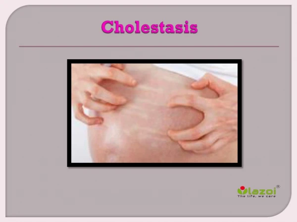
Definition of Cholestasis • Decrease in bile flow due to impaired secretion by hepatocytes or to obstruction of bile flow through intra-or extrahepatic bile ducts • Elevation of serum conjugated bilirubin • Indicates hepatobiliary dysfunction • Cholestatic jaundice affects approximately 1 in every 2,500 infants
Cholestasis Guideline Committee Recommendations • Any infant noted to be jaundiced at 2 weeks of age be evaluated for cholestasis with measurement of total and direct bilirubin • Breast-fed infants who can be reliably monitored and who have an otherwise normal history (no dark urine or light stools) and physical exam may be asked to return at 3 weeks • If jaundice persists, have measurement of total and direct bilirubin at that time
Causes of Cholestasis in Infant <2-months • Obstructive cholestasis: • Biliaryatresia: • Occurs in 1 in 10,000 to 20,000 infants • Obliteration or discontinuity of the extrahepatic biliary system, resulting in obstruction to bile flow • Cause is unknown • Important to diagnose BA early as ideal time for successful Kasai is 45-60 days
Obstructive Cholestasis • Alagille syndrome: • dominantly inherited disorder of variable expressivity. The gene has been identified as the Jagged1 (JAG1) • congenital cardiac defects (PPS) • posterior embryotoxon in the eye • dysmorphicfeaures • butterfly vertebrae. • Liver biopsy will show bile duct paucity • liver transplant for hepatic decompensation, bone fractures, pruritus, and xanthomas
Obstructive Cholestasis: • Choledochal Cyst • Can be diagnosed with ultrasound • Inspissated bile • Cystic fibrosis • Neonatal sclerosingcholangitis • Congenital hepatic fibrosis/Caroli’s disease
Hepatocellularcholestasis • Idiopathic neonatal giant cell hepatitis • Histologic appearance of widespread giant cell transformation • non-specific and may be associated with infectious, metabolic, and syndromic disorders • Needs close follow up and may self resolve • Infection • Sepsis • Cytomegalovirus, HIV, Toxoplamosis, Syphilis
Hepatocellularcholestasis • Genetic/metabolic disorders • a1-antitrypsin deficiency (A1AT) • Tyrosinemia • Galactosemia • Hypothyroidism • Progressive familial intrahepaticcholestasis (PFIC) • Cystic fibrosis • Panhypopituitarism • Toxic/secondary • Parenteralnutrition-associated cholestasis
Presentation: Questions to Ask on History • H/o Neonatal infection • UTI, sepsis and viral infection • Feeding history and history of weight gain • metabolic disease can cause anorexia, FTT, and jaundice • Bowel history • Vomiting - metabolic disease, pyloric stenosis, bowel obstruction • Delayed stooling—CF, hypothyroidism • Diarrhea—infection, metabolic disease, PFIC1, CF • Clay colored stool—biliaryobstruction • Dark urine color • Source of nutrition • Composition of formula: • Galactose containing galactosemia • Fructose or sucrose containing hereditary fructose intolerance
Presentation: Questions to Ask on History • Lethargy • Hypothyroidism, panhypopituitarism, sepsis, or infection • Excessive bleeding • coagulopathy, vitamin K deficiency • Similar problem with parents or among siblings • A1AT deficiency, Alagille syndrome, cystic fibrosis, PFIC • Consanguinity • Risk for autosomal recessive inheritance • Maternal infection that can affect baby • TORCH infections, occassionally HBV • Cholestasis of pregnancy • May be seen in PFIC • Past ABO or Rh disease, or Rh negative
Presentation: Physical Exam • Global assessment of general health and nutrition status • Dysmorphic findings (Alagille’s) • HEENT • Slit-lamp findings (Alagille’s , cataracts) • Chest/heart • Murmur or evidnece of congenital heart disease (biliaryatresia, Alagille’s) • Abdomen • Liver and spleen size and consistency • Distention, Ascites, Abdominal wall vasculature • Skin • Jaundice, bruising, petechiae, rashes • Neurologic • General assessment of vigor, tone, and symmetry • Diaper • Dark urine (conjugated hyperbilirubinemia) • Pale or clay colored stool (cholestasis, rule out obstruction)
Diagnosis: Labs • UA • Urine reducing substances • Thyroid function studies • Alpha-1-antitrypsin • Sweat chloride or mutation analysis for CF gene • Follow-up newborn screen • Total and direct bilirubin • LFT, GGT • Synthetic function • PT/PTT, glucose, albumin • CBC • Urine and blood culture • Viral serologies • TORCH infections, HBsAg, CMV, HIV if indicated
General Bilirubin Guidelines • If the TB is <5, an abnormal direct bilirubin is defined as a value >1mg/dL • If the TB is >5, an abnormal direct bilirubin is defined as a value that is >20% of the total • As the TB increases, the DB naturally increases • “Percentage system” is used when the TB reaches a threshold of 5 • DB may also vary greatly across labs, which may have practical implications
Diagnosis: Imaging Studies • Ultrasound with dopplers • Choledochal cyst • Portal vein thrombosis • For BiliaryAtresia • Triangular cord sign (TC) - obliterated fibrous ductal remnant in the portahepatis • May demonstrate absence of the gallbladder and no dilatation of the biliarytree • HIDA scan • Consider biliaryatresia if good hepatic uptake with no evidence of excretion into the bowel at 24 hours • Not specific for biliaryatresia • Pretreatment with phenobarbital (5 mg/kg/day for 5 days) to increase biliary secretion may help minimize false positives
If Concern for Biliary Obstruction • Refer to Hepatology • May need: • Liver biopsy • For BA portal tract fibrosis, edema, ductular proliferation, and cholestasis with the appearance of bile plugs • Intra-op cholangiogram • Further work up for cholestasis including directed genetic testing
Management of Cholestasis(NASPGHAN Guidelines) General algorithm for 2-8 week old infant
Management of Cholestasis • Management depends on underlying condition • General Management Principles • Optimize nutrition and growth • Fat soluble vitamin supplementation • Consider ursodiol to help with bile flow in obstructive processes • Routine physical exam and labs to assess for signs of progressing liver disease
Management of BiliaryAtresia • Kasai portoenterostomy • Excise extrahepaticbiliary tree • Portahepatis is transected at the level of the liver capsule to expose the remaining ductules • Jejunal Roux loop is anastomosed to the cut surface • Complications following Kasai • Ascending cholangitis. • Even with successful Kasai hepatic fibrosis or cirrhosis can develop • Portal hypertension • Liver transplantation • Consider if persistent or progressive cholestasis, cirrhosis with hepatic dysfunction, or portal hypertension with ascites and varicesunresponsive to management
Biliary Transport Defects • Three conditions comprise the currently known group of biliary transport defects • PFIC I: Mutations in the FIC1 gene (ATP8B1) • FIC1 mediates the flipping of aminophospholipids from outer to inner hemi-leaflet of the canalicular membrane • FIC1 is located on other tissues including the pancreas and intestine leading to other extrahepatic signs and symptoms • PFIC II have defects in the canalicular bile salt export pump (BSEP) caused by mutation in ABCB11 • BSEP is responsible for transporting bile acids from inside the hepatocyte into the bile canaliculus • PFIC III caused by mutations in ABCB4 • Encodes multidrug resistance-associated protein 3 (MDR3) and mediates the flopping of aminophospholipids from inner to outer hemi-leaflet of the canalicular lipid bi-layer • Liver disease in PFIC results from the effects of hepatocellular accumulation of bile acids
Biliary Transport Defects:Clinical Presentation & Diagnosis • Patients with forms of these diseases present with • Jaundice and/or pruritus • Life-threatening hemorrhage, secondary to cholestasis-related vitamin K deficiency • Patients with PFIC I and II have markedly elevated serum bile acid levels with mildly elevated serum bilirubin, normal serum gamma glutamyl transpeptidase (GGT) • PFIC III have high serum GGT levels • Definitive diagnosis of a specific form of PFIC is dependent upon identification of characteristic genetic defects
Biliary Transport Defects:Treatment and Outcomes • Supplement fat-soluble vitamins • Pruritis • Ursodeoxycholic acid, Cholestyramine, Rifampin, and opioid antagonists • Surgical interruption of the enterohepatic circulation of bile acids with either ileal bypass or diversion may be effective • Severe defects in BSEP are associated with a high risk of hepatocellular carcinoma • Severe defects in BSEP and MDR3 deficiency are typically associated with an unremitting form of cholestasis that are unresponsive to medical and surgical therapies necessitate liver transplantation • FIC1 is a systemic disease and the post-transplant course can be problematic including intractable diarrhea, liver steatosis leading to cirrhosis, and recurrent pancreatitis • Overall, with optimal surgical intervention the long-term prognosis for children with PFIC is excellent
Summary • It is essential to detect liver disease in an infant through a careful history, physical examination, and fractionation of the serum bilirubin • Biliary atresia is the most common disorder leading to cholestasis during the first postnatal months • Many intrahepatic disorders, such as congenital infection and inborn errors of metabolism can lead to cholestasis • Early recognition and diagnostic evaluation of the cholestatic infant is needed to manage the complications of the many metabolic and infectious liver diseases or surgery to relieve obstruction for biliaryatresia
References • Hartley, JL. BiliaryAtresia. Lancet 2009; 374: 1704-13 • Moyer V, Freese DK, Whitington, PF. Guideline for the Evaluation of Cholestatic Jaundice in Infants: Recommendations of the North American Society for Pediatric Gastroenterology, Hepatology and Nutrition. Journal of Pediatric Gastroenterology and Nutrition. 2004; 39:115–128 • Suchy, F. Neonatal Cholestasis. Pediatrics in Review. 2004; 25; 388-396. • Superina R, Magee JC, Brandt ML. The Anatomic Pattern of BiliaryAtresiaIdentfied at timeof Kasai Hepatoportoenterostomy and early postoperative clearance of jaundice are significant predictors of transplant free survival. Annals of Surgery 2001; 254: 577-585.