1 / 44

Hyaline cartildge in an area of the trachea

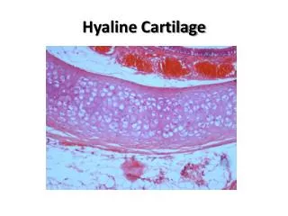
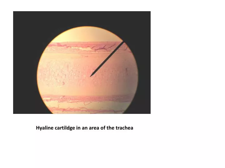
Hyaline cartildge in an area of the trachea. On high power the chondrocytes and lacunae are visible. View of compact bone on low power. Compact bone on medium power- structures of osteon are visible. Areolar tissue showing the two types of fibers and nuclei of the cell.

merton
Download Presentation

Hyaline cartildge in an area of the trachea

An Image/Link below is provided (as is) to download presentation Download Policy: Content on the Website is provided to you AS IS for your information and personal use and may not be sold / licensed / shared on other websites without getting consent from its author. Content is provided to you AS IS for your information and personal use only. Download presentation by click this link. While downloading, if for some reason you are not able to download a presentation, the publisher may have deleted the file from their server. During download, if you can't get a presentation, the file might be deleted by the publisher.

E N D

Presentation Transcript


  1. Hyaline cartildge in an area of the trachea

  2. On high power the chondrocytes and lacunae are visible

  3. View of compact bone on low power

  4. Compact bone on medium power- structures of osteon are visible

  5. Areolar tissue showing the two types of fibers and nuclei of the cell

  6. Dense regular fibrous CT on medium power

  7. Dense regular connective tissue- on high power the wavy organization of the tissue is visible

  8. Mesenchyme (embryonic)- tissue is pound of the periphery of the structure

  9. On high power- the cell nuclei and cytoplasmic extensions is mixed with the reticular fibres

  10. Adipose on medium power- as seen in the hypodermis

  11. Adipose tissue on high power

  12. Multipolar neuron- and associated glial cells

  13. 5 layers of the epidermis- 2 visible layers

  14. Layers of epidermis

  15. STRUCTURES OF DERMIS

  16. Hair follicle and surrounding dense fibrous irregular connective tissue

  17. Sebaceous Gland

  18. Arrector pili muscle

  19. Sudoriferous gland

  20. Suderiferous Gland

  21. Pacinian corpuscle

  22. Basal Cell carcinoma- medium power entire tumor is visible

  23. Basal Cell Carcinoma- on high power, individual cancer cells are visible

  24. BRAIN

  25. Dorsal (superior) view: look at lobes and structure of cerebrum and cerebellum

  26. Ventral (inferior) view: notice the trigeminal nerve arising from the pons

  27. Oculomotor nerves, optic chiasma and olfactory bulbs visible

  28. Midsaggital section

  29. A view of the posterior midbrain

  30. View of the Pineal gland

  31. Pituitary gland demo

  32. Grey and white matter demo

  33. Spinal cord model

  34. Retina Slides

  35. view of the optic disc and nerve

  36. Retina

  37. Clear view of three layers of retina and blood vessels in choroid

  38. Overall view of the three tunics

  39. One view of rods and cones

  40. Rods and cones (pointer on a cone)

  41. Spinal Cord Slides- Anterior (round) and Posterior (pointy) horns of grey matter

  42. Posterior root ganglion

  43. Posterior nerve root

  44. Anterior nerve root

More Related