Download
1 / 8
80 likes | 168 Views
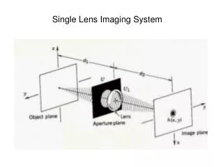
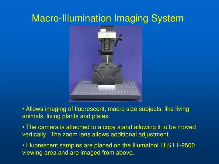
Macro-Illumination Imaging System. Allows imaging of fluorescent, macro size subjects, like living animals, living plants and plates. The camera is attached to a copy stand allowing it to be moved vertically. The zoom lens allows additional adjustment.

E N D
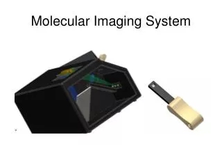
Macro-Illumination Imaging System • Allows imaging of fluorescent, macro size subjects, like living animals, living plants and plates. • The camera is attached to a copy stand allowing it to be moved vertically. The zoom lens allows additional adjustment. • Fluorescent samples are placed on the Illumatool TLS LT-9500 viewing area and are imaged from above.
Imaging Cabinet • This cabinet is used with the imaging system to provide a dark environment • It is useful to contain moving animals during the viewing process
LT-9500 Illunatool Tunable Lighting System • Excitation of fluorescent dyes, such as GFP, contained within the samples occurs via the above lighting model. • It consists of a lamp supply which delivers a single light source. It gets divided into two equal Epi line lights, which are placed above and to the side of the sample. • The result is a strong excitation signal that has little optical noise.
Sample mouse image • Red fluorescent Protein (RFP) imaging in brain of nude mouse
More sample images • Green Fluorescent Protein (GFP)-labeled tumor growing on colon of mouse.
More sample images • Another image of a GFP-labeled tumor
Sample image of a culture dish containing GFP transformed cells
GFP Plant Image Two cuttings of canola (Brassica napus). The one on the left is engineered with mGFP5-ER under the control of the CaMV 35S promoter. The one on the right is wild type.