Download
1 / 16
160 likes | 306 Views
How Do We Know What We Know? Neuroscience Methods. Tools for Viewing Brain Structure Neuroanatomy. Cell Body Staining Tract (Myelin) Staining Tract Tracing Immunocytochemistry. Tools for Viewing Brain Function Neurophysiology. Where can signals be recorded?.

E N D
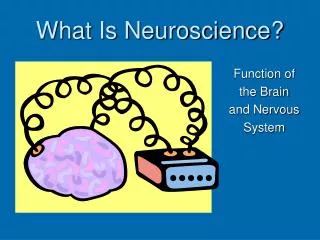
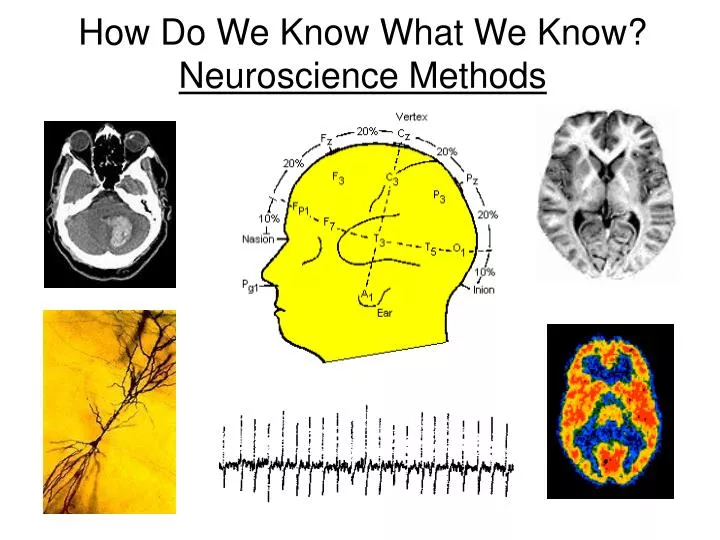
How Do We Know What We Know? Neuroscience Methods
Tools for Viewing Brain StructureNeuroanatomy • Cell Body Staining • Tract (Myelin) Staining • Tract Tracing • Immunocytochemistry
Tools for Viewing Brain FunctionNeurophysiology Where can signals be recorded? Channels Inside Outside Many “Patch Clamp” “Intracellular” “Extracellular” “Mass Unit”
Tools for Viewing Brain FunctionNeurophysiology Where can signals be recorded? 1 = EEG 2 = ECoG 3 = Intracranial 1 2 3 x x x
Tools for Viewing Brain FunctionNeurophysiology Where can signals be recorded? • Patch Clamp Recording • Intracellular Recording • Extracellular Recording • Mass Unit Recording • Evoked Potentials • Electrocorticography (ECoG) • Electroencephalography (EEG)
Electroencephalogram (EEG) Emotiv Demo
Brain Imaging • Computed Tomography Scan (CT Scan) • Positron Emission Tomography (PET) • Magnetic Resonance Imaging (MRI) • Functional Magnetic Resonance Imaging (fMRI)
Brain Imaging CT scan • Also called a CAT scan • Computerized axial tomography • X-ray of brain tissue • Shows brain structure
Tools for Viewing Brain Structure and Activity PET scan • Positron Emissions Tomography • Patients drinks radioactive glucose and image shows areas of brain activity.
Tools for Viewing Brain Structure and Activity MRI • Magnetic Resonance Imaging • Exposes brain to magnetic field • Shows brain structure
Tools for Viewing Brain Structure and Activity fMRI • Functional MRI • Uses magnetic field • Detects blood flow changes • Not harmful • Shows brain structure and activity
Behavioral Neuroscience • Stereotaxic Surgery • Lesion Production/Injury • Electrical Brain Stimulation • Microinjection
Injury – Clinical Findings Paul Broca and “Tan”
Injury – Clinical Findings Phineas Gage
Brain Mapping Finding out what each part of the brain does Electrical Brain Stimulation Also: Transcranial Magnetic Stimulation