Download

1 / 45
450 likes | 462 Views
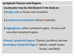
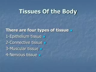
The Lymphatic System Lecture Outline Understanding of lymph tissues Dynamic of lymphoid tissues Distribution of Lymphoid tissues.

E N D
The Lymphatic System Lecture Outline Understanding of lymph tissues Dynamic of lymphoid tissues Distribution of Lymphoid tissues
Skin=2m2Respiratory= 100m2Gut =400mVascular tree= 100000kmVelocity of lymphocyte in main vessel=0.5m/secondVelocity of lymphocyte in o.vessel=0.5mm/secondtotal lymphoid cell in blood is 1x10¹¹ total lymphoid cell in stationary is 9x10¹¹ Principles of Human Anatomy and Physiology, 11e
Introduction • The Lymphatic SYSTEM STRUCTURE • Immunophysiology of Lymphatic S • Development of Lymphatic tissues Principles of Human Anatomy and Physiology, 11e
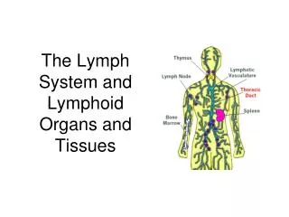
Lymphatic System • Organs, vessels and a fluid called lymph • similar to interstitial fluid • Organs involved • red bone marrow • thymus • spleen • lymph nodes • diffuse lymphatic tissue • tonsils, adenoids & peyers patches Principles of Human Anatomy and Physiology, 11e
Skin=2m2Respiratory= 100m2Gut =400mVascular tree= 100000kmVelocity of lymphocyte in main vessel=0.5m/secondVelocity of lymphocyte in o.vessel=0.5mm/secondtotal lymphoid cell in blood is 1x10¹¹ total lymphoid cell in stationary is 9x10¹¹ Principles of Human Anatomy and Physiology, 11e
Thymus Gland Figure 22.5 • Large organ in infants (70 g) but atrophied as adult (3 g) • 2 lobed organ located in mediastinum • Capsule & trabeculae divideit into lobules • Each lobule has cortex &medulla • Cortex • tightly packed lymphocytes ¯ophages • Medulla • reticular epithelial cells produces thymic hormones • Hassall’s corpuscles Principles of Human Anatomy and Physiology, 11e
Thymus Gland • Large organ in infants (70 g) but atrophied as adult (3 g) • 2 lobed organ located in mediastinum • Capsule & trabeculae divideit into lobules • Each lobule has cortex &medulla • Cortex • tightly packed lymphocytes ¯ophages • Medulla • reticular epithelial cells produces thymic hormones • Hassall’s corpuscles Principles of Human Anatomy and Physiology, 11e
Thymus Gland • Large organ in infants (70 g) but atrophied as adult (3 g) • 2 lobed organ located in mediastinum • Capsule & trabeculae divideit into lobules • Each lobule has cortex &medulla • Cortex • tightly packed lymphocytes ¯ophages • Medulla • reticular epithelial cells produces thymic hormones • Hassall’s corpuscles Principles of Human Anatomy and Physiology, 11e
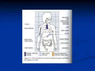
Lymphatic Nodules • Concentrations of lymphatic tissue not surrounded by a capsule scattered throughout connective tissue of mucous membranes • mucosa-associated lymphoid tissue (MALT) • Peyer’s patches in the ileum of the small intestine • Appendix • Tonsils form ring at top of throat - Figure 23.2 • adenoids (pharyngeal tonsil) • palatine tonsils (on each side wall) • lingual tonsil in the back of the tongue Principles of Human Anatomy and Physiology, 11e
Lymph Nodes - Overview • Lymph nodes are encapsulated oval structures located along lymphatic vessels (Figures 22.1a and 22.6). • They contain T cells, macrophages, follicular dendritic cells, and B cells. • Lymph enters nodes through afferent lymphatic vessels, is filtered to remove damaged cells and microorganisms, and exits through efferent lymphatic vessels. • Foreign substances filtered by the lymph nodes are trapped by nodal reticular fibers. • Macrophages then destroy some foreign substances by phagocytosis and lymphocytes bring about the destruction of others by immune responses. • Lymph nodes are the site of proliferation of plasma cells and T cells. • Knowledge of the location of the lymph nodes and the direction of lymph flow is important in the diagnosis and prognosis of the spread of cancer by metastasis; many cancer cells are spread by way of the lymphatic system, producing clusters of tumor cells where they lodge. (Clinical Application) Principles of Human Anatomy and Physiology, 11e
Lymph Nodes • Flow is in one direction • afferent vessels lead in • sinuses lead to efferent vessels that exit at hilus • Only nodes filter lymph Principles of Human Anatomy and Physiology, 11e
Spleen Figure 22.7 • 5 inch organ between stomach & diaphragm • Hilus contains blood & lymphatic vessels • Stroma consists of capsule, trabeculae, fibers & fibroblasts • Parenchyma consists of white pulp and red pulp • white is lymphatic tissue (lymphocytes & macrophages) around branches of splenic artery • red pulp is venous sinuses filled with blood & splenic tissue (splenic cords) Principles of Human Anatomy and Physiology, 11e
Circulation of Lymphatic system Principles of Human Anatomy and Physiology, 11e
Functions of the Lymphatic System • Draining excess interstitial fluid & plasma proteins from tissue spaces • Transporting dietary lipids & vitamins from GI tract to the blood • Facilitating immune responses • recognize microbes or abnormal cells & responding by killing them directly or secreting antibodies that cause their destruction Principles of Human Anatomy and Physiology, 11e
Lymphatic Vessels and Lymph Circulation • Lymphatic vessels begin as blind-ended lymph capillaries in tissue spaces between cells (Figure 22.2). • Interstitial fluid drains into lymphatic capillaries, thus forming lymph. • Lymph capillaries merge to form larger vessels, called lymphaticvessels, which convey lymph into and out of structures called lymph nodes (Figure 22.1). Principles of Human Anatomy and Physiology, 11e
Lymphatic Vessels & Circulation • Capillaries that begin asclosed-ended tubes foundin spaces between cells • Combine to form lymphaticvessels • resemble veins with thinwalls & more valves • Fluid flows through lymph nodes towards large veins above the heart • lymph emptied into bloodstream Principles of Human Anatomy and Physiology, 11e
Lymphatic Capillaries • Lymphatic capillaries have a slightly larger diameter than blood capillaries and have overlapping endothelial cells which work as one-way valves for fluid to enter the lymphatic capillary. • Anchoring filaments attach endothelial cells to surround tissue (Figure 22.2). • A lymphatic capillary in the villus of the small intestine is the lacteal. It functions to transport digested fats from the small intestine into blood. Principles of Human Anatomy and Physiology, 11e
Lymphatic Capillaries • Found throughout thebody except in Avasculartissue (cartilage, epidermis& cornea) • Structure is designed to lettissue fluid in but not out • anchoring filaments keep tubefrom collapsing under outside pressure • overlapping endothelial cells open when tissue pressure is high (one-way valve) Principles of Human Anatomy and Physiology, 11e
Lymph Trunk and Ducts • The principal lymph trunks, formed from the exiting vessels of lymph nodes, are the lumbar, intestinal, bronchomediastinal, subclavian, and jugular trunks (Figure 22.3). • The thoracic duct begins as a dilation called the cisterna chyli (Figure 22.4) and is the main collecting duct of the lymphatic system. • The thoracic duct receives lymph from the left side of the head, neck, and chest, the left upper extremity, and the entire body below the ribs. • It drains lymph into venous blood via the left subclavian vein. Principles of Human Anatomy and Physiology, 11e
Lymph Trunks & Ducts • Vessels unite to form trunks & thoracic ducts • Right side head, arm & chest empty into right lymphatic duct and rest of body empties into thoracic duct • Lymph is dumped directly into left & right subclavian veins Principles of Human Anatomy and Physiology, 11e
Immunophysiology of Lymphatic system Lymphatic System Transport by lymphatic system Lymphatic circulation Mechanism of filtration Principles of Human Anatomy and Physiology, 11e
Lymphatic System Thin wall in all tissues Intersitial fluid Thorasic duct Dynamic of fluid Starling hypothesis Drinker hypothesis Selection hypothesis Principles of Human Anatomy and Physiology, 11e
Transport by lymphatic systemFatty acids labelled with radioactive carbona-short fatty acids—intestine--blood--liver—Metabolizeb-long fatty acids– intestine—lymphHormones from tissues to blood Principles of Human Anatomy and Physiology, 11e
Lymphatic circulationcapillary (thin)----Vessel Elastic fibersysterna chylii transport 500cc /hour Principles of Human Anatomy and Physiology, 11e
Mechanism of filtration1-Pumping action of lymphatic vessels2- = effect tissues motion3- = action of terminal lymphatic capillary4-the pressure of fluid in the tissues space Principles of Human Anatomy and Physiology, 11e
Lymphatic traffic( In normal condition)1-Lymph circulate through gut associated lymph tissues---blood---PP----mesenteric lymph node-----Thorasic duct --- heart2-Skin---peripheral LN----Central LN---thorasic duct---heart3-Recirculate through lymphoid organ LN---LNLymphocyte reticulate through spleenblood spleen bloodspleen--- miner ---lymph4-lymphocyte recirculation through to thorasic duct Principles of Human Anatomy and Physiology, 11e
Lymphatic traffic( In abnormal condition)1-Lymphocyte circulate through gut associated---lesion-- lymph tissues---blood---PP----mesenteric lymph node-----Thorasic duct --- heart2-Lymphocyte crculate through Skin---infalammation---peripheral LN----Central LN---thorasic duct---heart3-Recirculate through lymphoid organ—infammation in organ--- LN---LN Principles of Human Anatomy and Physiology, 11e
Kinetic of flow in L.N Principles of Human Anatomy and Physiology, 11e
Development Lymphatic tissues • Emberyonic • adults Principles of Human Anatomy and Physiology, 11e
DEVELOPMENT OF THE LYMPH TISSUES • Lymphatic vessels develop from lymph sacs, which develop from veins. Thus, they are derived from mesoderm. • Lymph nodes develop from lymph sacs that become invaded by mesenchymal cells (Figure 22.8). Principles of Human Anatomy and Physiology, 11e
Developmental Anatomy • Begins to develop by 5thweek • Lymphatic vessels developfrom lymphatic sacs thatarise from veins • Jugular sac & cisterna chyliform thoracic duct • Sacs develop into lymph nodes • Spleen develops in gastric mesentery • Thymus is outgrowth of 3rd pharyngeal pouch Principles of Human Anatomy and Physiology, 11e