Download
1 / 106
2.08k likes | 3.06k Views
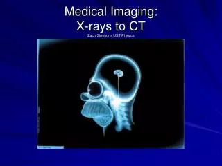
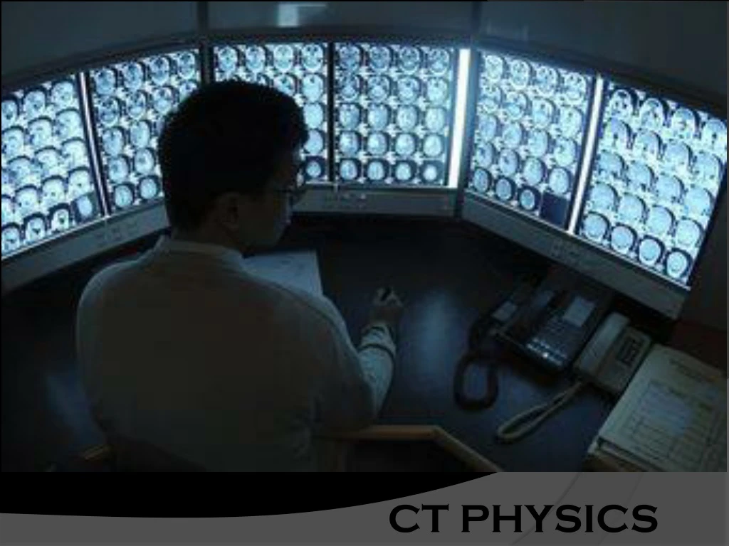
CT PHYSICS. Computed Tomography “T omos in G reek means sections ”. Computed tomogragphy ( CT) is a process of creating a cross -sectional tomographic plane or slice of any part of the body in which computer is used to make a mathematical reconstruction of a tomograph (CT image).

E N D
Computed tomogragphy (CT) is a process of creating a cross -sectional tomographic plane or slice of any part of the body in which computer is used to make a mathematical reconstruction of a tomograph (CT image) Definition
History When was the x ray machine invented? • Before x ray machines were invented, broken bones, tumors and the location of bullets were all diagnosed by physical examination and a doctor's best guess. • Patients paid the price of these approaches. • Then on November 8th of 1895, a German physics professor Wilhelm Conrad Roentgen made a remarkable discovery.
History • was a Professor at University in Germany. • Working with a cathode-ray tube in his laboratory, Roentgen observed a fluorescent glow of crystals on a table near his tube.
History • He concluded that a new type of ray was being emitted from the tube. • This ray was capable of passing through the heavy paper covering and exciting the phosphorescent materials in the room. • He found that the new ray could pass through most substances casting shadows of solid objects.
History • Roentgen also discovered that the ray could pass through the tissue of humans, but not bones and metal objects. • One of Roentgen's first experiments late in 1895 was a film of the hand of his wife, Bertha.
History • In June 1896, only 6 months after Roentgen announced his discovery, X-rays were being used by battlefield physicians to locate bullets in wounded soldiers.
1963: Alan Cormack formulated X-ray absorption in tissues & developed theory of image reconstruction. 1972: Sir Godfrey Hounsfield. History :
X-Ray image formation– 2D with super- imposition of tissues Conventional Tomography – 2D without superimposition of tissues but with better resolution and accuracy INTRODUCTION
Computed Tomography Scan is trans-axial or transverse scanning with image plane perpendicular to long axis of the body .
CT scan revolutionized diagnostic imaging. • When compared to conventional radiography. • Better contrast resolution • No superimposition of tissues. • Less scatter radiation • 3D imaging • Combined with radionuclide studies
BASIC PRINCIPLE:-THE INTERNAL STRUCTURES OF AN OBJECT CAN BE RECONSTRUCTED FROM MULTIPLE PROJECTIONS OF THE OBJECT • Well collimated X-ray beam is directed to the patient. • Attenuated image forming radiation. • Measured by detector • Reconstruction of the image by mathematical equations by computer and displays on monitor.
Basic Principle : • Pencil like or fan shaped x-ray beam used. • Source and detector move around synchronously around the object of interest
GENERATIONS • “GENERATIONS": The Stages of Evolution of Equipment Systems Used in CT Scanning. • Progressively decreased the Scanning Time.
FIRST GENERATION SCANNERS • Translate –rotate motion. • Pencil shaped beam . • Single detector used.
EMI SCANNER • Hounsfield 1972. • Only for brain evaluation. • Linear and rotatory gantry motions. • After 1 translation, tube and detector rotate 1 degree.
Linear motion was repeated 160 times. Axis of rotation passed through the center of head. 1 degree rotation between each of these linear movements.
Scan time for each pair of tomographic sections: 4.5 to 5 min • Total scan time for a clinical study 25 minutes. • CT image was reconstructed
SECOND GENERATION • Head & body scan. • Fan shaped beam.Narrow fan beam (30-100) • Multiple detectors – 5 to 30. • Linear and rotatory movements. • 30 sec scan time
Fewer linear movements are needed as there are more detectors to gather the data. Between linear movements, the gantry rotated 30 degree. Only 6 times the linear movements got repeated
DISADVANTAGES • Increased scatter radiation due to fan shaped beam. • Increased intensity towards edges of the beam because of body shape. Bow tie filter used - to reduce the intensity towards the edges of the beam because of body shape
THIRD GENERATION • Rotate(tube)-Rotate(detectors) : Linear motion is completely eliminated. • Pulsed wide fan beam 500 – 550 • Detectors are perfectly aligned with the X-Ray tube • Scan time< 5secs
Single image is computed from more than 1000 projections. X ray tube is continuously on. Number of lines in each projection is equal to number of detectors. THIRD GENERATION
Advantages • A constant source to detector path length maintained. 1.Good image reconstruction 2.Better x-ray beam collimation
FOURTH GENERATION : • Continuous wide fan beam(500-550) • Ring of detectors(> 2000) • Rotate(tube)-Fixed(detector) • X-ray tube rotates in a circle inside the detector ring • Scan time< 2 secs
ADVANTAGES: Free from ring artifacts DISADVANTAGES: Higher patient dose Higher cost
System components • The gantry: x-ray tube, detector array, high voltage generator, patient support couch, mechanical supports. • The computer: Image reconstruction • The operating console: imaging control and image viewing
X-RAY TUBES FOR HEAT DISSIPATION- • High speed rotating anode is used. • Large heat loading( Upto 8M HU) capacity.
SCINTILLATION CRYSTALS “A scintillating crystal is any material that produce light as a result of external influence, particularly ionizing radiation ” • Used by all rotate fixed ( fourth generation) and some rotate rotate units. (third generation). Difference b/w scintillation crystals and intensifying screens ?
The X-ray tube rotates continuously in one direction,whilst the table on which the patient is lying is mechanically moved through the X-ray beam.