Download
1 / 19
200 likes | 233 Views
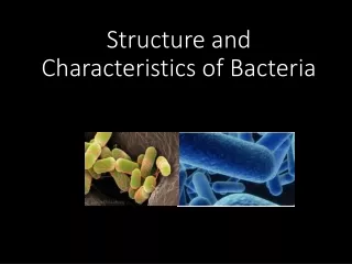
Morphology and structure of bacteria. Oral Microbiology for dentistry MUDr. Lenka Č ernohorsk á, Ph.D. Size of bacteria. Pathogenic bacteria: mainly around 1 – 5 μm (1 μm = 10 -3 mm) Staphylococcus: the diameter circa 1 μm

E N D
Morphology and structure of bacteria Oral Microbiology for dentistry MUDr. Lenka Černohorská, Ph.D.
Size of bacteria Pathogenic bacteria: mainly around 1 – 5 μm (1 μm = 10-3 mm) Staphylococcus: the diameter circa 1 μm Even smaller: rickettsiae (circa 0.5 μm) chlamydiae (elementary bodies circa 0.3 μm) mycoplasmas (circa 0.2 – 0.25 μm )
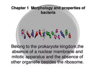
Various shapes of bacteria • Cocci (spherical) • Rods /bacilli: can be short long robust, thin • Spirochaete (helical)
Common bacterial forms • Cocci in chains – streptococci • Cocci in clusters – staphylococci • Cocci in pairs – diplococci (neisseria) • Bacilli in chains • Curved bacilli • Spore-bearing bacilli
Structures II. Cytoplasm- is enclosed within the cell membrane, contains organelles.Here occur most cellular activities and metabolic pathways. The part of the cytoplasm that is not held within organelles is called the cytosol (a gel, with a network of fibers dispersed through water) Capsule– gelatinous layer around the bacterium is composed of polysaccharides, proteins – inhibits phagocytosis, helps the adhesion, is used in preparation of vaccines Fimbriae – hair-like filaments, mediate adhesion to receptors Inclusions– serve as sources of stored energy (polysaccharides)
Gram-staining (more in practical lessons) • Crystal violet – 30 s • Iodine solution – 30 s • Alcohol – decolorize until violet is removed • Safranin – 60 s G- pink groups G+ purple groups
Cell wall Results: • different in G- and G+ bacteria • principle of Gram staining is still unknown, but it is used for hundred years • G+: Crystal violet attaches to peptidoglycan (PG) than arise complex with Iodine, complex is not washed by alcohol, that is why the final result is purple. • G-: Little amount of PG inside a cell wall, a little complex/no complex? is washed by alcohol, Safranin is needed for visualisation.
Gram-positives Staphylococcus Streptococcus Bacillus Clostridium Listeria Corynebacterium Yeasts Gram-negatives Escherichia Salmonella Haemophilus Pseudomonas Mycoplasma Examples ofG+andG–microbes
Flagella • filaments composed of flagellin • responsible for movement of bacteria May be located: (cocci and bacilli) • at one end:monotrichous, a single flagellum lophotrichous – many flagella • all over the outer surface – peritrichous • Spirochaets move by using the axial filament – produce undulation motion
Sporulation (bacterial winter sleep) • In bad conditions some kind of bacteria sporulate • Spora contains a high concentration of calcium dipicolinate, is resistant to heat, radiation, chemicals, dehydratation, it can remain for many years • Better conditions- spore transform itself into reproducing bacterial cell again.
Sporulation process septum
Spores - facts • Spore contains: DNA, small amount of cytoplasm, cell membrane, peptidoglykan, very little water, thick keratin-like coat (with calcium dipicolinate)
Relevance of bacterial spores • Resistance to heat and chemicals! • They cannot be easily achieved by boiling • Other methods of sterilisation like autoclaving should be used • So for ex. Bacillus stearothermophilus is used for evaluation of the sterilisation efficacy of autoclaves!
Types of spores 1, 4 – central 2, 3, 5 – terminal 6 - lateral 2 – with inclusions 3, 4, 5, 6 – spores deforming a bacterial cell 1,2 – spores not deforming
Spores terminal subterminal central
Thank you for your attention More in your textbooks.