Download
1 / 24
290 likes | 546 Views
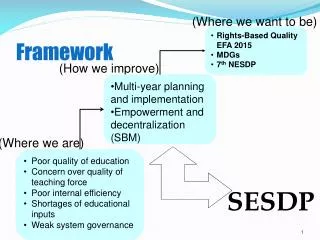
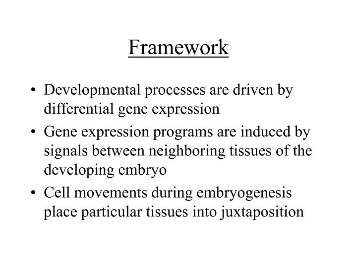
Framework. Developmental processes are driven by differential gene expression Gene expression programs are induced by signals between neighboring tissues of the developing embryo Cell movements during embryogenesis place particular tissues into juxtaposition. “Official glossary” (from Wolpert).

E N D
Framework • Developmental processes are driven by differential gene expression • Gene expression programs are induced by signals between neighboring tissues of the developing embryo • Cell movements during embryogenesis place particular tissues into juxtaposition
“Official glossary” (from Wolpert) • Morphogen: Any substance active in pattern formation whose spatial concentration varies and to which cells respond differently at different levels • Morphogenesis: The process involved in bringing about changes in form in the developing embryo • Pattern formation: Process by which cells in a developing embryo acquire identities that lead to a well-ordered spatial pattern of activities
What development accomplishes • Differentiation of all of the required cell-types from a single fertilized egg (oocyte) • Morphogenesis: Precise arrangement of these cells into tissues and organs • Pattern formation: Precise arrangement of tissues and organs to achieve a reproducibly working organism capable of reproduction • Epigenesis: the de novo formation of an organism from “disordered” egg cytoplasm
Demonstration of nuclear potential in Acetabularia (1930’s) Figure 2.5 page 31 Gilbert
The nucleus and Epigenesis • The nucleus contains the instructions that drive epigenesis/development • Chromatin is the instructional unit (DNA plus proteins). The state of the chromatin is set by “epigenetic control” mechanisms • Covert “epigenetic” changes occur during the early cleavages of the fertilized egg into the blastomeres of the embryo
Embryonic cell division is not the same in all kinds of organisms Next slide So there is a sense that these zygotes “know what they are” when the begin to divide. The yolk and its position obviously plays a role in determining the cleavage pattern. But, there are sometimes other products stored in the egg by the mother that act as morphogenetic determinants.
Major morphogenetic strategies • Autonomous specification • Morphogenetic determinants deposited in the egg become segregated by cell division • Determination of cell fate is early • Conditional specification • Cell fate determination is later and depends on the position of the blastomere in the embryo • Removal of cells is compensated for by others • Each cell has the potential to give rise to more cells than it normally does
Pages 56-66 of text Specification of cell fate: Autonomous vs. Conditional
Syncitial specification • Mainly seen in insects (Drosophila) • Gradients form, over time, of morphogens deposited in the egg by the mother • These morphogens become segregated by cell membranes which grow into the egg • Drosophila lectures: End of February
GASTRULATION • The process that puts cells into position to have their fates determined • Gastrulation involves a particular repertoire of cell movements which can be classified • Gastrulation will result in the formation of three “germ layers” from which the organs of the embryo will arise
Gastrulation and Conditional Specification • Newly positioned tissues (germ layers) interact with one another to “induce” organ formation • Cell-cell interactions via receptors (juxtacrine) • Soluble signaling molecules (paracrine) • Morphogen gradients link position to cell fate • Signal transduction activates gene expression which leads to specification and lineage commitment
Morphogen gradients: different concentrations of a factor induce different gene expression
Stages of commitment to cell fate • Specification • Changes in gene expression which are labile and changeable. The gene expression “allows” that cell to differentiate along a pathway but does not irreversibly commit the cell • Determination • Further changes in gene expression which seal the lineage fate of the cell and eliminate alternative choices
Pro-B cells from Pax-5 deficient mice are “specified” IL-7 Im syk Oct-2 l5 btk E2A VpreB blk PU.1 B29 lyn EBF “Pro-B cell”
Pro-B cells from Pax-5 deficient mice are “specified”but not “determined” T cell mouse Im syk Oct-2 l5 btk E2A VpreB blk PU.1 B29 lyn EBF Macrophage M-CSF “Pro-B cell” Trance Dendritic cell Osteoclast IL-2 GM-CSF NK cell
Recent data illustrating the concepts of specification vs. determination Nutt, SL, et. al. (1999) Commitment to the B-lymphoid lineage depends on the transcription factor Pax-5 Nature, 401:556-562 E2A EBF Pax-5 B lymphocyte development committed specified
Forming a solid organ • How do cells “stick together” to form tissue? • Coordination of tissues from multiple germ layers to form a single functional organ • Tissue layers • Organ polarity • Cell adhesion molecules: Cadherins (p66-74)
Cadherins (Ca2+dependent adhesion molecules) See also Website 3.8
Types of cadherins • E- cadherin: expressed on early embryonic cells in mammals. Later becomes restricted to embryonic and adult epithelial tissue • P-cadherin: Trophoblast cells (placental) • N-cadherin: First mesodermal, later CNS • EP-cadherin: frog blastomere adhesion • Protocadherins: not connected to catenin
Mechanisms of cadherin based cell “sorting” into tissues • Differential expression of cadherin type • Neural vs epidermal cells (N vs. E cadherin) • Different levels of cadherin expression • Oocyte positioning in follicle • Loss or switch of cadherin expression • Neural crest emigration from the neural tube • Protocadherin switching in frog gastrulation • See pages 311-312 (Chapter 10 of Gilbert)