Download
1 / 52
600 likes | 974 Views
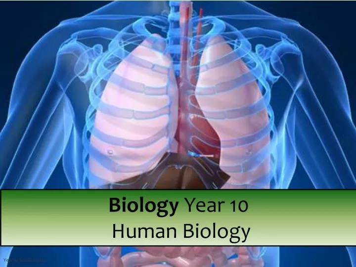
Biology Year 10 Human Biology. The structure and functions of the human (as an animal) systems: skeletal, digestive, circulatory, respiratory.

E N D
Biology Year 10 Human Biology Year 10 Science 2012
The structure and functions of the human (as an animal) systems: skeletal, digestive, circulatory, respiratory. All vertebrates share the same basic body plan, with tissues and organs functioning in a similar manner. We focus on the human body, studying structure (anatomy) and function (physiology). Year 10 Science 2012
The structure and functions of the human (as an animal) systems: skeletal, digestive, circulatory, respiratory. Animals are made of complex systems of cells, which must be able to perform all of life’s processes and work in a coordinated fashion to maintain homeostasis (a stable internal environment). During a human’s early development, groups of cells specialize into three fundamental embryonic layers. These embryonic layers differentiate into a number of specialized cells and tissues. Tissues are groups of cells similar in structure and function . Year 10 Science 2012
The structure and functions of the human (as an animal) systems: skeletal, digestive, circulatory, respiratory. Different tissues functioning together for a common purpose are called organs (eg, stomach, kidney, lung, heart). Organ systems are composed of individual organs working together to accomplish a coordinated activity. For example, the heart, veins and arteries all play a role in circulation. Year 10 Science 2012
The Human skeletal system. The human skeletal system is an internal skeleton that serves as a framework for the body. This framework consists of many individual bones and cartilages. This also includes ligaments and the tendons which hold bones to each other and to muscles that contract and cause the bones to move. The adult human skeleton contains more than 200 bones. The skeleton also provides mechanical protection for many of the body's internal organs, reducing risk of injury to them, storage of minerals and production of blood cells. Year 10 Science 2012
The main bones in the Human skeletal system. The main bones of the human skeleton are divided into two groups. The axial skeleton is the central group of bones based around the spinal (vertebral) column. The appendicular skeleton consists of bones or groups of bones which “hang” off the axial skeleton.
The Human skeletal system. The main bones of the human skeleton are: The Skull Shoulder girdle - clavicle and scapula Arm- humerus, radius and ulna Hand - Carpals, Metacarpals and Phalanges Chest- Sternum and Ribs Spine- vertebrae, Sacrum (5 fused or stuck together bones) and Coccyx (the tiny bit at the bottom of the spine). Pelvic girdle - Ilium, Pubis and Ischium. Leg- Femur, Tibia and Fibula Ankle- Talus and calcaneus Foot - Tarsals, Metatarsals and Phalanges
A joint occurs where two bones meet . Movement of the skeleton occurs at the joints where two or more bones meet. There are different categories of joints. Freely movable, or synovial, joints allow a range of movement determined by the structure of the joint. Examples of movable joints are the ball and socket (shoulder), hinge (elbow), pivot (between the skull and neck) and ellipsoidaljoint (hand and arm). Ligamentsare inelastic connective tissues which hold bones together in a joint.
The structure of the skeleton and muscles of the human arm The muscles are attached to the bones by tendons. When the muscles contract they shorten and move the bones at the joints. All of the bones in the skeleton require muscles in order to move them. The muscles are attached by the nervous system to the brain which controls their movement – either voluntary or automatically when required. Year 10 Science 2012
The antagonistic muscles and bones act together to flex or extend the arm Muscles can contract, but are not designed to actively lengthen, so they are arranged as opposing antagonistic pairs. As one muscle shortens, another is stretched and vice versa. The contacting muscles move the bones they are attached to.
The digestive system The digestive system converts foods to simple substances that can be absorbed and used by the cells of the body. It is composed of the mouth, pharynx, oesophagus, stomach, small intestine and large intestine and is aided by several accessory organs (liver, gall bladder, and pancreas). Year 10 Science 2012
Digestion breaks large molecules of food into small ones, which can then pass through the wall of the gut into the blood. Ingestion How we take in food using our mouths Digestion Breaking food into smaller pieces Absorption Taking food into the blood Egestion Removal of food from the anus anus 12 Year 10 Science 2012
Ingestion is how we take in food using our mouths. • Canine and incisors teeth rip, tear and bite pieces of the food. • Suitable sized pieces of food enter the mouth. • Pre-molars and molars grind the food into smaller pieces. • Saliva is mixed with the food from the saliva glands. • Enzymes in the saliva start breaking down (digesting) the food. • Lumps of chewed food are swallowed down the esophagus. • Food moves into the stomach Year 10 Science 2012 SJ Gaze
The internal structure of a human tooth. The tooth has a hard covering of enamel which protects it and gives it strength to bite and chew food. When tooth decay occurs the enamel is eaten away by bacteria and tooth pain occurs because acid and infection reach the dentine and tooth nerves. The tooth is embedded into the jaw bone by the roots which secure it. Year 10 Science 2012
How the different types of human teeth are used when eating. There are 32 teeth in a full set of adult teeth. The incisors at the front of the mouth are used to bite pieces of food. The bicuspids (also called premolars) and the molars at the back of the mouth are used to chew food. The canines are bigger and used to tear food. Year 10 Science 2012
How the different types of Animal (carnivores) teeth are used when eating. Tiger carnivorous cat Alligator carnivorous reptile Otter carnivorous fish eater Sharp pointed canines Year 10 Science 2012 SJ Gaze
How the different types of Animal (Herbivores) teeth are used when eating. Horse grazing herbivore Rabbit grazing rodent Grey kangaroo Australian grazing marsupial Chisel-like incisors No canines Grinding molars SJ Gaze
How the different types of Animal (omnivores) teeth are used when eating. Badger eats more animals than plants. Human eats more plants than animals Flattened molars to grind plants Canines much smaller Year 10 Science 2012 SJ Gaze
How the different types of Animal teeth (specialist) are used when eating. Baleen whales The specialized filter-feeding mechanism of baleen whales enables them to feed low on the food chain by primarily eating zooplankton and schooling fishes. Giant Anteater sucks up ants with long tongue and has no need for teeth at all. SJ Gaze
The gut is a coiled tube and is the site of digestion and absorption. The second stage in food processing is digestion DefinitionDigestion Breaking food into smaller pieces Once food is ingested it moves down into the oesophagus and then into the stomach DefinitionOesophagus The tube that food travels from the mouth to the stomach through DefinitionStomach Organ from the digestive system that digests food Year 10 Science 2012 SJ Gaze
The gut is a coiled tube and is the site of digestion and absorption. The third stage in food processing is absorption Definition Absorption Taking food into the blood This absorption occurs in the small intestine which the food moves into from the stomach and water absorption occurs in the large intestine DefinitionSmall Intestine Organ from the digestive system that absorbs food into the blood Blood containing the absorbed food then moves to the liver to be further processed Definition Large intestine Organ from the digestive system that absorbs water from waste food DefinitionLiver Organ from the digestive system that stores and distributes food SJ Gaze
The main structure of the digestive system and its associated organs -oesophagus. The oesophagusis like a stretchy pipe that's about 25 centimeters long. It moves food from the back of your throat to your stomach. When you swallow a small ball of mushed-up food or liquids, a special flap called the epiglottiscloses over the opening of your windpipe to make sure the food enters the oesophagusand not the windpipe. Once food has entered the oesophagus muscles in the walls move in a wavy way to slowly squeeze the food through the oesophagus (peristalsis) and into the stomach. Year 10 Science 2012
The main structure of the digestive system and its associated organs – stomach. Your stomach is attached to the end of the oesophagus. It has three important functions: >to store the food you've eaten >to break down the food into a liquid mixture >to slowly empty that liquid mixture into the small intestine The stomach mixes, churns and mashes together all the small pieces of food into smaller and smaller pieces – called digestion. It does this with help from the strong muscles in the walls of the stomach and gastricjuicesthat also come from the stomach's walls. In addition to breaking down food, gastric juices also help kill bacteria that might be in the eaten food. Year 10 Science 2012
The main structure of the digestive system and its associated organs - small and large intestine. The small intestineis a long tube around 3.5 to 5 centimeters around, which connects from beneath your stomach and is about 7 meters long. The small intestine breaks down the food mixture even more so your body can absorb all the vitamins, minerals, proteins, carbohydrates, and fats. Food may spend as long as 4 hours in the small intestine and will become a very thin, watery mixture which can pass from the intestine into the blood. Small intestine
The small intestine is the site of absorption of the products of digestion. The walls of the small intestine are covered in protruding villi which increase the surface area and provide close contact for capillaries to absorb small food particles through the wall.
The main structure of the digestive system and its associated organs - small and large intestine. The large intestineis about 7 to 10 centimeters, which is wider than the small intestine from which it joins on. It would measure about 1.5 meters long spread out. Most of the nutrients have already been removed from the food mixture before it enters but there is waste and water left over. Before it leaves the large intestine, it passes through the part called the colon where the body is able to absorb the water and some minerals into the blood. Year 10 Science 2012
The main structure of the digestive system and its associated organs – Pancreas, gall bladder and liver. These organs send different juices to the first part of the small intestine. These juices help to digest food and allow the body to absorb nutrients. The pancreas makes enzymes that help the body digest fats and protein. Enzymes from the liver called bile helps to absorb fats into the bloodstream. The gallbladder serves as a warehouse for bile, storing it until the body needs it.
The main structure of the digestive system and its associated organs – pancreas, gall bladder and liver. The nutrient-rich blood comes directly to the liver from the small intestine for processing. The liver filters out harmful substances or wastes, turning some of the waste into more bile. The liver sorts howmanynutrients will be distributed to the body, and how many will stay behind in storage. The liver stores certain vitamins and a type of sugar your body uses for energy. Year 10 Science 2012
The main structure of the digestive system and its associated organs – rectum and anus. The final stage of food processing is egestion, where the waste food that hasn’t been absorbed is moved through the rectum and out of the body through the Anus. Definition Egestion Removal of wastes from the anus Definition Anus part of the digestive system through which wastes exit the body Year 10 Science 2012
The food we eat is divided into four main groups There are four main food groups Carbohydrates >supply instant energy >include sugar and starches >are supplied by fruit (sugars) or by cereals (starches) Lipids >act as energy stores >include fats and oils >are energy rich >are made of fatty acids Proteins >are used for growth and repair >are found in meat and eggs >are made of amino acid chains Minerals and Vitamins >body needs them in small amounts >found in many foods >non-organic substances SJ Gaze
We use tests to determine which type of food is present. Lipids – present in fats and oils Protein – present in meat and eggs Some of the food rubbed into filter paper. Remove food and allow to dry Sodium hydroxide solution Add benedicts solution Mixture turns purple if protein present Broken up food particles A ‘grease spot’ is left where the food was rubbed in if lipid was present Year 10 Science 2012 SJ Gaze
We use tests to determine which type of food is present. Starch –present in cereals Glucose – type of sugar Add Benedict’s solution Heat mixture gently Place a few drops of iodine on the food Mixture turns orange if glucose present Broken up food particles in water Food will turn black if starch is present Year 10 Science 2012 SJ Gaze
The circulatory system The structure of the circulatory system consists of blood, blood vessels and the heart. The function of the circulatory system is to be the body’s transportation system, moving oxygen, carbon dioxide, nutrients, wastes, hormones, vitamins, minerals and water throughout the body. It also aids in regulation of temperature. Year 10 Science 2012
The external and internal structure of the mammalian heart and its function. The mammal has a four chambered heart with full division between the ventricles. This means there is no mixing of the oxygenated and deoxygenated blood. It contains valves, and heart beat is controlled by the nervous system pacemaker. Year 10 Science 2012
The external and internal structure of the mammalian heart and its function. The function of the Circulatory system must: >ensure delivery of blood to all tissues >adapt so that blood flow can be controlled and changed to individual tissues or the body as a whole >convert a pulsating blood flow in the arteries into a steady flow in the capillaries to allow optimum diffusion to and from the cells >return blood to the heart Year 10 Science 2012
Arteries carry blood away from the heart, veins carry blood towards the heart. The vascular system is composed of arteries, arterioles, capillaries, venules and veins. The structure of the vessels in the different parts of the vascular system varies and the differences relate directly to the function of each type of vessel.
Arteries carry blood away from the heart, veins carry blood towards the heart. There are three main types of blood vessels: >arteries, which carry blood away from the heart at relatively high pressure. >veins, which carry blood back to the heart at relatively low pressure. >capillaries which provide the link between the arterial and venous blood vessels Veinshave thinner, non-muscular walls to accommodate the lower blood pressure of the blood returning to the heart. Arterieshave thick muscular and elastic walls to accommodate the high blood pressure of the blood leaving the heart. Capillaries are only one cell thick.
Arteries carry blood away from the heart, veins carry blood towards the heart. The Arteries and veins have different structures because they have different functions Year 10 Science 2012
Arteries carry blood away from the heart, veins carry blood towards the heart. Circulatory system A Deoxygenated blood enters the right atrium of the heart via the vena cava.B Deoxygenated blood is pumped from the right ventricle, via the pulmonary artery, to the lungs. C In the lungs, blood picks up oxygen and gets rid of carbon dioxide.D Oxygenated blood travels back to the heart in the pulmonary vein.E Oxygenated blood is pumped to vital organs and the whole body from the left ventricle, via the aorta.F Digested food is absorbed by blood through the walls of the small intestine.G Digested food is processed and stored in the liver.H Waste products (urea, water, salts etc.) are removed from the blood in the kidneys.I Blood supplies the rest of the body with food and oxygen. It carries away waste products.
Capillaries link arteries with veins and are the sites of exchange with the tissues. Arteries divide into smaller arterioles around targeted tissues. The arterioles divide once more into smaller capillaries which are only one cell thick. Nutrients and O2 diffuse across the membranes of the capillaries from the blood to the cell supplied – from high to low concentration. Waste products, such as CO2, will diffuse from the cell into the capillary near the venule end. The venulesrejoininto veins and waste product contained within the blood will be pumped back to the heart. Year 10 Science 2012
Blood consists of fluid (serum) containing cells – white blood cells, red blood cells and platelets. White blood cells (leucocytes) – some produce antibodies to tag foreign and harmful objects, others surround and eat tagged objects. White blood cells prevent infection becoming established. Make up the immunity system. Cells contain nucleus. Red Blood cells (erythrocytes) – contain haemoglobin, a pigment which binds to oxygen molecules and carries it through the circulatory system. They contain no nucleus. Platelets – they collect where there is a hole in the blood vessels and stop the unwanted flow of blood by clotting. Year 10 Science 2012
Red blood cells carry oxygen, attached to haemoglobin, around the body of a mammal. Mammalian red blood cells (erythrocytes) dispose their nuclei and cell organelles when they are mature in order to provide more space for haemoglobin (oxygen carrying pigment). Because the cells have no nucleus or organelles, they cannot produce any RNA, and therefore they cannot divide or repair themselves. They are constantly replaced by new red blood cells. Red blood cells are biconcave disks and this shape optimises the cell for the exchange of oxygen in the lungs. The red blood cells are flexible so they can fit through tiny capillaries, where they release their oxygen to cells. Year 10 Science 2012
Plasma transports glucose, carbon dioxide, urea and hormones. Plasma carries dissolved molecules required by the body to each cell and waste products like carbon dioxide and urea out of the body. Plasma also carries a large number of important proteins. Albumin, the main protein in blood, helps control the water content of tissues and blood. Plasma is usually yellow in colour due but can become milky when it transports fat absorbed from the intestines to other organs of the body. Year 10 Science 2012
The respiratory system The respiratory system consists of organs that deliver oxygen to the circulatory system for transport to all body cells. The respiratory system also assists in removing the waste product of carbon dioxide from the body. Oxygen is a vital element for metabolism. The exchange of oxygen and carbon dioxide between body cells, blood and the air in the lungs is called respiration. Year 10 Science 2012
The structure of the mammalian breathing system The human respiratory system consists of the nose, pharynx, larynx, trachea, bronchi, and lungs (composed of bronchioles and alveoli). The lungs are housed in an airtight pleural cavity framed by the rib cage and diaphragm. Year 10 Science 2012
The structure of the mammalian breathing system Air (comprised of about 21% oxygen) enters the nose, where tiny hairs filter out dust and particles. Tissues moisten and warm the air, making it more suitable for gas exchange in the lungs. Air passes from the pharynx to the larynx (containing vocal cords) and into the trachea. Year 10 Science 2012
The structure of the mammalian breathing system The trachea divides into the left and right bronchi which subdivide into smaller and smaller tubes called bronchioles. These airways are lined by mucous membranes and many cilia hairs which trap and remove particles from the lungs. Bronchioles open into the alveoli which are clustered like grapes. Year 10 Science 2012
The structure of the alveoli and blood capillaries enable gaseous exchange to occur. Only one cell thick, alveoli have direct contact with capillaries for gas exchange. The grapelike arrangement of alveoli creates an enormous surface area sufficient for exchanging enough oxygen and carbon dioxide for the entire body. Year 10 Science 2012
The importance of diffusion in gaseous exchange across the alveoli. Oxygen is transported to the cells of the body mainly by binding to haemoglobin which is found within red blood cells. Carbon dioxide is transported by diffusion into the plasma. Diffusion occurs because the oxygen is in higher concentration in the alveoli and moves to the lower concentration in the blood of the capillary. The CO2 in the blood diffuses into the alveoli and out of the body because it also moves from high to low concentration. Oxygen enters into alveoli from bronchioles The oxygen molecules must diffuse through both the lining of the alveolus and the lining of the blood capillary and is eventually picked up by red blood cells Year 10 Science 2012
Air breathed out contains more carbon dioxide and less oxygen than air breathed in. Breathing (the movement of air in and out of the lungs) is produced by pressure differences created by changing the size of the pleural cavity. The diaphragm contracts and the rib cage is raised. The volume of the pleural cavity is increased, creating a partial vacuum between the lung cavity and the atmosphere, and air enters the lung. Year 10 Science 2012