Download
1 / 31
330 likes | 548 Views
Hazards of Prematurity. Fred Hill, MA, RRT. Introduction. The major factor of morbidity and mortality in the neonate is the degree to which the organ systems have not yet developed. The earlier in gestation that birth occurs, the higher the degree of morbidity and mortality. Introduction.

E N D
Hazards of Prematurity Fred Hill, MA, RRT
Introduction • The major factor of morbidity and mortality in the neonate is the degree to which the organ systems have not yet developed. • The earlier in gestation that birth occurs, the higher the degree of morbidity and mortality.
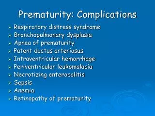
Introduction • Some of the complications experienced by premature infants include : - respiratory distress syndrome (RDS) - bronchopulmonary dysplasia (BPD) - retinopathy of prematurity (ROP) - necrotizing enterocolitis (NEC) - intraventricular hemorrhage (IVH) • pulmonary interstitial emphysema (PIE) • apnea
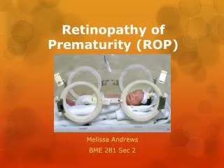
Retinopathy of Prematurity (ROP) • Formerly retrolental fibroplasia (RLF) • Early seen that oxygen usage led to RLF • With ↓ use of O2,, ↑ CP & ↑ mortality • Factors contributing to ROP • ↑ O2 levels • Retinovascular immaturity • Circulatory & respiratory instability
Incidence (ROP) • 25 – 35% of premature infants (<36 weeks) • 5-10 % have at least stage 3 • 3-5 % result in blindness
Pathophysiology (ROP) • ↑ PaO2 → retinal vasoconstriction • Retinal vessel necrosis (vaso-obliteration) • Surviving blood vessels proliferate • Proliferation into vitreous • Vessels in vitreous hemorrhage • Hemorrhages form scar tissue • Retinal detachment & blindness
Many Factors Involved (ROP) Immaturity, hyperoxia, hypoxia, blood transfusions, IVH, apnea, infection, hypercapnia, PDA, prostaglandin synthetase inhibitors, vitamin E deficiency, lactic acidosis, bright lighting, early intubation, hypotension, NEC
Treatment (ROP) • Vitamin E modest benefit • Cryotherapy: A probe that has been cooled to –20 C with nitrous oxide is introduced behind the eye and the avascular portion of the retina is frozen preventing further abnormal vessel proliferation. • Laser therapy: the avascular portion of the retina is photocoagulated. • Surgical interventions: such as vitrectomy and lensectomy.
Prevention (ROP) • Cautious use of oxygen in premature infants • Usually PaO2 50-80 mm Hg
Bronchopulmonary Dysplasia (BPD) - Incidence • Follows treatment for RDS • Primary etiologic factors • High pressures of ventilation • High FIO2 • Prematurity • Other contributing factors • Presence of PDA • Fluid overload • Exact etiology unknown
Definition (BPD) • Radiographic features as described by Northway et al (1967) • Oxygen dependence past 28 days following mechanical ventilation • Oxygen dependence beyond 36 weeks without mechanical ventilation (also neonatal chronic lung disease – NCLD or Wilson-Mikity Syndrome)
Radiographic Staging of BPD • Stage I (first 3 days): bilateral ground glass (RDS) • Stage II (3-10 days): opaque with obscuring of cardiac shadow • Stage III (10-20 days): multiple cyst formation; visible cardiac shadow • Stage IV (>28 days): ↑ lung density and formation of large, irregular cysts
Radiographic Staging of BPD • Stage I (first 3 days): bilateral ground glass (RDS) • Stage II (3-10 days): opaque with obscuring of cardiac shadow • Stage III (10-20 days): multiple cyst formation; visible cardiac shadow • Stage IV (>28 days): ↑ lung density and formation of large, irregular cysts
Mechanical ventilation Small ET tube with small leak High frequency ventilation Humidification of inspired gases CPT: PRN, not on regular schedule Careful aseptic suctioning Corticosteroids Aerosolized bronchodilators Theophyllline Fluid therapy/diuretics Digoxin Nutrition Vitamin E (inconclusive) Prevention: low pressure, low FIO2 Treatment (BPD)
Pulmonary Interstitial Emphysema (PIE) • PIE occurs when air dissects throughout the interstitial tissue of the lungs. • It results from the chronic use of high PEEP, peak inspiratory pressures and prolonged inspiratory times • PIE is classified into: • Intrapulmonary interstitial pneumatosis (when air remains within the lung tissue) • Intrapleural pneumatosis: extra alveolar air is confined by the visceral pleura, forming blebs.
Pulmonary Interstitial Emphysema (PIE) • As the air dissects and collects in the interstitium, the small airways and vessels are compressed. • V-Q mismatches follow and lead to a deterioration in blood gases • High ventilator pressures required to correct the worsening blood gases, cause more air leak into the interstitium and V-Q mismatch worsens again.
Hyper aerated lung fields in a 27 week infant with PIE Pulmonary Interstitial Emphysema (PIE)
Shortly before the death of this infant, lungs remain hyperaerated. Pulmonary Interstitial Emphysema (PIE)
Treatment of PIE • PIE is best treated by prevention • Low ventilation pressures may help avoid the onset of PIE • Selective intubation of the unaffected lung may allow injured lung to heal.
Intraventricular Hemorrhage (IVH) Factors • Immaturity • Fluctuating cerebral blood flow Mechanical Ventilation Effects • ↑ MAP may retard blood return • Variations in PaO2 & PaCO2 may induce cerebral vasoconstriction &/or vasodilation
Necrotizing Enterocolitis (NEC) Definition • Ischemia & necrosis of the intestine Etiology • Mucosal wall injury • Bacterial invasion • Formula feeding (breast milk seems to be protective)
Blood in stool (guaiac positive) Abdominal distension Lethargy Poor feeding tolerance/residuals Vomiting Apnea Temperature instability DIC Necrotizing Enterocolitis (NEC) Signs
Necrotizing Enterocolitis (NEC) Treatment • Prevention: good handwashing • Stop oral feedings • NG suction • Antibiotics (ampicillin & gentamicin) • Abdominal X-rays • Increased FIO2 • Surgical intervention following perforation
Necrotizing Enterocolitis (NEC) X-Ray Findings • Pneumatosis intestinalis (gas trapped in intestinal wall) • Portal vein gas • Free air in peritoneum – usually indicates perforation
Apnea • Periodic breathing: Short, recurring pauses in spontaneous ventilation of 5 to 10 seconds duration – considered normal in premature infant • Clinical definition of apnea: Complete cessation of breathing for a period of 20 seconds or any pause in breathing accompanied by bradycardia, ↓ in SpO2, and/or color change • Definition of apnea in adult sleep apnea: repeated epidsodes of complete cessation of airflow for 10 seconds or longer
Causes of Apnea • Periodic breathing: Short, recurring pauses in spontaneous ventilation of 5 to 10 seconds duration – considered normal in premature infant • Clinical definition of apnea: Complete cessation of breathing for a period of 20 seconds or any pause in breathing accompanied by bradycardia, ↓ in SpO2, and/or color change • Definition of apnea in adult sleep apnea: repeated epidsodes of complete cessation of airflow for 10 seconds or longer
Respiratory: RDS, congenital upper airway anomalies, airway obstruction, postextubation, CPAP, pneumonia, hypoxia Cardiovascular: CHF, PDA, anemia, polycythemia, tachycardia & bradycardia, sepsis, polycythemia CNS: IVH, meningitis, seizures, sedation, kernicterus, immaturity of respiratory centers, tumors GI: NEC, gastroesophageal reflux, stooling Metabolic: Hypoglycemia, hypo- & hypernatremia, hypocalcemia, hypo- & hyperthermia Enviromental: inappropriate temperature, suctioning, feeding See Table 10-5 Causes of Apnea
Treatment • Identify and correct underlying problem • CPAP • Oxygen • Mechanical ventilation • Drugs • Mehtylxanthines: theophylline, aminophylline, caffeine • Doxapram • Apnea monitors