Download
1 / 45
470 likes | 1.03k Views
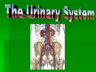
The Urinary System. Functions of the Urinary System. Provide control over plasma/body electrolyte & water balance Maintains plasma osmolarity Maintains plasma pH Excrete nitrogenous waste (urea & creatine) Nitrogenous wastes often generated by amino acid metabolism

E N D
Functions of the Urinary System • Provide control over plasma/body electrolyte & water balance • Maintains plasma osmolarity • Maintains plasma pH • Excrete nitrogenous waste (urea & creatine) • Nitrogenous wastes often generated by amino acid metabolism • Excrete drugs & other metabolites • Urinary drug tests
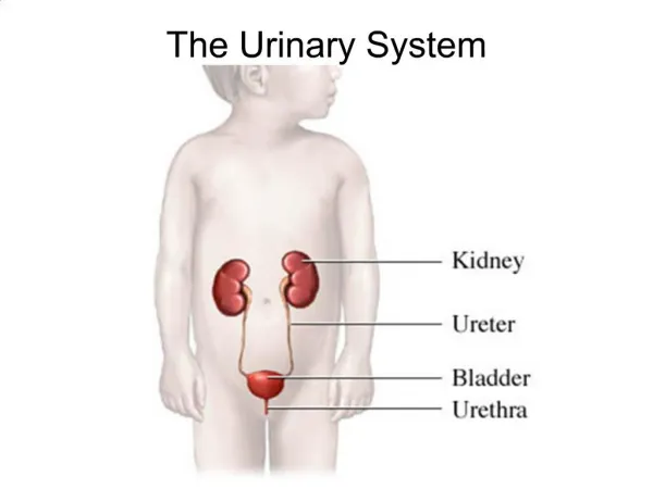
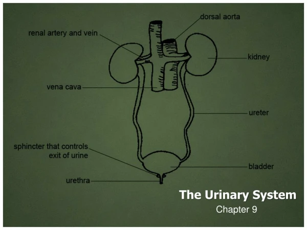
Anatomical Location • Posterior abdominal cavity • Retroperitoneal (behind the peritoneum) • Right kidney usually 1.5-2cm caudal than left • Kidneys connected to bladder via ureters • Bladder located at anterior base of the pelvis
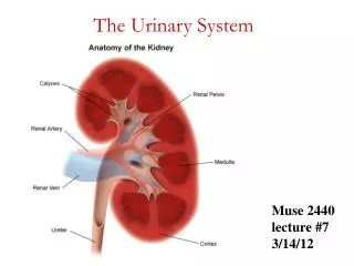
Gross Renal Anatomy • Kidneys covered by “capsule” • Intimate to kidney = Fibrous capsule • Semi-transparent fibrous connective tissue • Can be seen on male cadaver as a white layer • Layer of adipose connective tissue • Renal fascia that anchors kidney to abdominal wall & peritoneum
Gross Renal Anatomy • Bisect the kidney and you see some key gross anatomical features: • Outer cortex (red due to dense capillary network) • Inner medulla region (dark red due to tubules & vasculature) • Usually 8-15 renal pyramids separated by renal columns • Apex of each renal pyramid = renal papillae (acts like a funnel) • Renal papillae anastomize into minor calyx • Minor calyces anastomize into major calyx
Microscopic Renal Anatomy • Microscopically, the nephron is the functional unit of the kidney • Over 1 million per kidney • Knowledge of bloodflow pattern through kidney is very helpful in understanding kidney function • Renal artery – interlobar arteries – arcuate arteries – interlobular arteries – afferent glomerular arterioles • Afferent glomerular arterioles carry blood to the initial region of the nephron (glomerulus) • Remember the gradient of pressure: highest in artery, lowest in capillary • Arterioles retain enough pressure to permit the “filtration” to occur
Nephron • Glomerulus is a very complicated structure whose architecture is vital for kidney filtration • Capillary bed is squished into a ball (glomerulus) • Capillary bed is fenestrated (large windows, but not as big as sinusoids in liver) • Capillary is “encased” by unique cells = “podocytes” (foot cells) • Podocytes extend “pedicels” onto the capillary body • Between each foot or pedicel = “filtration space” where plasma contents can filter through glomerulus into the Bowman’s capsule (glomerular capsule)
The podocyte / pedicel structure is akin to the astrocytes in the blood – brain barrier. The podocytes & astrocytes wrap themselves around the capillary bed in their respective region. In the nephron, the podocytes allow MORE plasma to filter through than the astrocytes in the brain.
Nephron • Once plasma has filtered out of the glomerulus, it enters the Bowman’s capsule (glomerular capsule) • A funnel-like structure that initiates the nephron tubule network • Into the proximal convoluted tubule • Simple cuboidal epithelium with a distinctive apical brush border/microvillus structure • Region of solute re-absorption • Into the loop of Henle (nephron loop) • Then “up” the distal convoluted tubule • Simple cuboidal epithelium with markedly less apical brush border structure • Region of water re-absorption • This loop structure, which places the proximal tubule in very close approximation to the distal tubule = countercurrent exchange mechanism
URINE Idealized tubule
Afferent arteriole PCT DCT glomerulus Active transport: electrolytes, glucose, proteins etc. “osmotic drag” of water Efferent arteriole Water-diluted blood, now iso-osmotic “Concentrated” (plasma has been filtered out at glomerulus) Slightly hypertonic, “extra-concentrated” Vasa recta (tubule capillary network)
Ureter • Urine flows from distal convoluted tubule – collecting duct – renal papilla – minor calyx – major calyx – renal pelvis – ureter • Series of “collecting funnels” • From renal pelvis –posterolateral edge of the bladder • 3 layers of tissue (markedly similar to intestinal tract) • Inner muscosa (forms continuous “sheet” from renal tubule – bladder) • Known as “transitional epithelium”…can stretch • Middle muscularis (opposite of the GI tract) • Innermost layer (intimate to mucosa) = longitudinal muscle • Outermost layer = circular muscle • Outer Adventitia
Bladder • Posterior to pubic symphysis • 4 layer organ • Internal mucosa (transitional epithelium like ureter) • Folded into “urinary bladder rugae” in order to allow bladder distension • Region of the trigone (urethral opening) does not have rugae • Submucosa • Muscularis • 3 layers of smooth muscle, similar to stomach • “detrusor muscle” = combined 3 layers • Base of trigone = neck = internal urinary/urethral sphincter • Adventitia (actually parietal peritoneum)
Urethra • Tube similar to ureter • Lined with urethral glands that secrete mucus in order to protect lining • Short distance between internal urinary/urethral sphincter (smooth muscle) and external urinary/urethral sphincter • Note that the external urinary sphincter is NOT at the very end of the urethra • Also recall how external urinary sphincter = skeletal muscle of the pelvic diaphragm • Actual constrictor muscle group = bulbous spongiosum
Urethra • Male vs female slightly different • Female urethra short and relatively “straight” • Urethral orifice located between labia minora • A single-use tube (only for excretion of urine) • Short & straight = more prone to infection (similar to estuchian canal of the middle ear) • Male urethra longer and “S” shaped, divided into 3 regions: • Prostatic: region passing through prostate gland • Receives prostate excretions & ejaculatory fluid • Membranous: region that passes through pelvic diaphragm • Location of the external urinary sphincter • Spongy: longest portion, to external urethral orifice • Where bulbourethral glands (Cowper’s glands) secrete pre-ejaculate lubricant into urethra • A DOUBLE-USE tube: urine & reproduction/ejaculate
Urination / Micturation • Reflex action to expel stored urine • In infants/young, this is a simple reflex • Voluntary control not developed until 2-3 years • Requires development of cerebral cortex & maturation of the spinal cord (recall the cauda equina) • Bladder can normally hold 1-1.5 liters of urine! • 200-300 ml urine is enough to trigger the distension reflex that tells you that it’s time • Stretch receptors trigger the micturation reflex • PNS triggers detrusor musclecontraction & internal urinary sphincter relaxation • Pudental nerve controls external urinary sphincter (bulbous spongiosum)
Kidney transplants • Kidney transplants are the most frequently transplanted organs in the world. • 12,000 kidney transplant procedures per year in USA alone. • Donor kidneys can be harvested from two sources: • Living donor (identical twin, serotype match) • Cadaver
Why Transplant a Kidney? • Indications for kidney transplant include: • Diabetes, glomerulonephritis, polycystic kidney disease, pyelonephritis. • End goals of the transplant include: • Improved quality of life, reduce patient care costs and strain on healthcare provider. • Reduce/remove the need for dialysis or CAPD (Continuous Ambulatory Peritoneal Dialysis). • Remove dietary restrictions.
Kidney Transplant Procedure • Identify acceptable patient (one that will tolerate transplant surgery) • Identify donor organ (living donor or cadaver). • Prepare patient (council, prophylactic immunosuppressant drugs, etc.) www.viscom.ohiou.edu/kring/home.html Donor is placed on their side to permit access to the donor kidney.
Kidney Transplant Procedure • Surgical removal of donor kidney (living donor). www.viscom.ohiou.edu/kring/home.html Primary incision (non-laparoscope)
Kidney Transplant Procedure • Surgical removal of donor kidney (living donor). • Locate and prepare kidney for removal. www.viscom.ohiou.edu/kring/home.html Entering the kidney capsid (connective tissue anchoring the kidney to the dorsal abdomen)
Kidney Transplant Procedure • Surgical removal of donor kidney (living donor). • Locate and prepare kidney for removal. www.viscom.ohiou.edu/kring/home.html Ligating renal vasculature and ureter prior to removal
Kidney Transplant Procedure • Surgical removal of donor kidney (living donor). • Locate and prepare kidney for removal. www.kidneytransplant.org/live-donorlaparoscopictransplant.html All attempts are made to include as much vasculature and ureter as possible.
Kidney Transplant Procedure • Surgical removal of donor kidney (living donor). • Locate and prepare kidney for removal. • Prepare donor kidney for serotype analysis (to minimize threat of rejection). www.viscom.ohiou.edu/kring/home.html Flushing donor kidney with ice cold saline prior to serotyping
Kidney Transplant Procedure • Surgical removal of donor kidney (living donor). • Locate and prepare kidney for removal. • Prepare donor kidney for serotype analysis (to minimize threat of rejection). www.viscom.ohiou.edu/kring/home.html Flushing donor kidney with ice cold saline prior to serotyping
Kidney Transplant Procedure • Surgical removal of donor kidney (living donor). • Locate and prepare kidney for removal. • Prepare donor kidney for serotype analysis (to minimize threat of rejection). www.viscom.ohiou.edu/kring/home.html Donor organs are kept on ice in order to minimize tissue degradation.
Kidney Transplant Procedure • Prepare recipient for donor organ (once serotyping has indicated a match). www.viscom.ohiou.edu/kring/home.html Contrary to donor procedure, recipient is supine to expose the pelvis.
Kidney Transplant Procedure • Prepare recipient for donor organ (once serotyping has indicated a match). • Donor kidney is inserted deep into the pelvic region, and all vessels are reconnected. www.kidneytransplant.org/patientguide/insurgrey.html Note: recipient’s kidneys are NOT removed.
Nephrolithiasis (Kidney Stones) • Kidney stones are hard pellets formed in the nephron as minerals crystallize in the tubule. • Calcium oxalate stones ( 70-80%) • Uric acid ( 7%) • Magnesium ammonium phosphate ( 2%) • Other (xanthine, cystine) ( 2%)
Nephrolithiasis (Kidney Stones) • If permitted to nucleate large enough, stones can lodge in the lumen of nephron and lead to urinary blockade, infection, nephritic damage, and severe pain. • 70-80% of the crystallized minerals from which stones can nucleate are actually passed out in urine without being noticed.
Stone Formation • Stones are formed when urine is supersaturated with the salts that are normally found in urine. • Volume reduction (low urinary volume) • High salt concentration (defective re-absorption by the tubular cells, excessive salt concentration in serum leading to excess salt in the glomerular filtrate). • Under supersaturated conditions, the salts will precipitate out and form crystals.
Stone Formation • Stone nucleation upon crystal formation can result from: • Decreased “protective” factors: • Mg2+, citrate, pyrophosphate etc. • Factors act to prevent crystal formation, or coat the crystal to prevent adherence to tubule wall. • Altered urinary pH: • pH < 5.5 can favor uric acid and cystine stone formation. • pH > 6 can favor calcium phosphate and magnesium ammonium phosphate stone formation.
Calcium Oxalate Stones • General treatment calls for: • Increased fluid intake • Increased calcium intake • Decreased oxalate intake • Spinach, strawberries, nuts, chocolate, tea, dark cola, wheat bran,
Uric Acid Stones • Treatment by: • Restricted protein diet (uric acid is derived from purine metabolism, and purine is found in protein from meat, beer, legumes, spinach, asparagus etc.). • Increase fluid intake
Magnesium Ammonium Phosphate Stones (Struvite) • Normally linked to urinary tract infection. • Treatments: • Antibiotic therapy (Lithostat to inhibit bacterial activity) • Urinary tract irrigation with organic acid (Struvite stones are acid soluble) • Recent evidence for “nanobacteria” as the major cause • Nano because they are smaller than 0.2m (cannot be “sterile filtered”)
Cystine Stones • Excessive cystine in urine (cystinurea) linked to genetic defect in the intestinal processing of cystine. • Cystine is soluble in urine at low pH, therefore treatments are aimed at acidifying urine. • Alkaline-Ash diet: • Milk, Fat (Nuts), Vegetables (except corn and lentils), Fruit (except cranberries, prunes, plums), Sweets: Molasses
Polycystic Kidney Disease (PKD) • Genetic disease resulting in gradual kidney degeneration due to cyst formation within the nephon • Etiology is still being studied • Linked to defective transporters along the nephron & absence of primary cilium among others
Renal Dialysis • Should urinary system fail, renal dialysis is an option • Either mechanical or through the use of patients own peritoneal cavity
Renal Dialysis • Chronic ambulatory peritoneal dialysis (CAPD) uses the patients peritoneal membrane as the filtration medium • Introduce dialysis fluid into the peritoneum, withdraw when it has “exchanged” and is full of metabolic waste (4-6 times daily)