Download
1 / 19
420 likes | 977 Views
Histology of Digestive Tract. Dr. Nabil Khouri. General Structure of Digestive Tract. Common Characteristics: Hollow tube composed of a lumen whose diameter varies. Surrounded by a wall made up of 4 principal layers : Mucosa

E N D
Histology of Digestive Tract Dr. Nabil Khouri
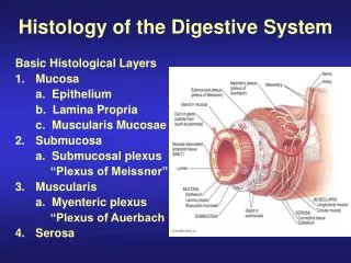
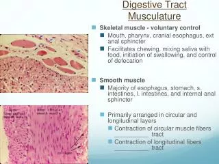
General Structure of Digestive Tract Common Characteristics: • Hollow tube composed of a lumen whose diameter varies. • Surrounded by a wall made up of 4 principal layers: • Mucosa • Epithelial lining; A lamina propria of loose connective tissues rich in blood, lymph vessels and smooth muscle cells; Muscularis mucosae. • Submucosa • Dense connective tissues with many blood and lymph vessels. • Muscularis • Contains smooth muscle cells, divide into 2 layers; internal (circular); external (longitudinal) • Serosa • Thin layer of loose connective tissue rich in blood and lymph vessels and adipose and single squamous covering epithelium (mesothelium)
Basic Mucosal Forms in GI . Protective mucosa – • Found in oral cavity, pharynx, esophagus, anal canal • Type – stratified sqaumous epithelium. . Secretory mucosa – • Found only in stomach • Type – glandular epithelium (packed tubular), Simple branched. . Absorptive mucosa – • Found in the Entire small intestine • Arranged in a fingerlike projection manner – villi • Duodenum only – sub-mucosal mucus Brunner’s glands – crypts - extend into the muscularis mucosa . Absorptive/protective mucosa – • Found in large intestine • Type – glandular epithelium (packed tubular), Strait, cells specialized for water and mucus absorption - Goblet cells
Histology of the Stomach Mucosa • Epithelial lining is composed of • Simple columnar, cells with round or oval nucleus at the base with granules • Goblet cells that produce a coat of alkaline mucus • Mucosa is folded to form gastric pits • Gastric pits (GP) containing gastric glands (Open into the bottom of GP) that secrete: • Gastric glands secrete gastric juice • Mucus – protects stomach wall from pepsin • Pepsin – splits protein • Hydrochloric acid – needed for pepsin to work • Intrinsic factor – aids in absorption of vitamin B12
Microscopic Anatomy of the Small Intestine • Structural modifications of the small intestine wall increase surface area • Plicae circulares: Characteristics of the jejunum • Transverse folds of the intestinal lining (deep circular folds of the mucosa and submucosa) semi lunar, Circular or spiral form • Villi: • fingerlike extensions of the mucosa (epithelium and lamina propria) • Microvilli: • tiny projections of absorptive mucosal cells’ plasma membranes • Lacteals • Terminal lymphatic in villus • Intestinal glands • Lined by entero-endocrine, goblet and stem cells
The Intestinal Villi • Central lacteal (lymphatic vessel) • Core capillaries • Core of connective tissue • Epithelial cells – Tall columnar with striated border Simple columnar epithelium Produces Mucin Enterocytes Tall columnar cells W microvilli form the brush border Lymphatic vessels - Paneth cells found at the base of the crypt
Small Intestine: Histology of the Wall • The epithelium of the mucosa is made up of: • Absorptive cells and goblet cells • Interspersed T cells (intraepithelial lymphocytes), and • Entero-endocrine cells • Intestinal crypts cells secrete intestinal juice • Peyer’s patches are found in the submucosa • Whereas, Brunner’s glands are found only in the duodenum and secrete alkaline mucus
Mucosa (M)-Villous form Glands- Coiled and branched Crypts of LieberKuhn. Brunner glands Tall columnar cells. Basally located nucleus Secret alkaline mucus Lysozyme and EG Factor Ducts pass through the MM and openinto the crypts Muscularis mucosa (MM) Extends between the glands Duodenum V ucosa M G
Mucosa of the Small Intestine • The mucosal surface is folded into villi • Increases surface area for absorption • Interior contains blood vessels and a lacteal • The mucosa and submucosa are thrown into circular folds Plicae Circulares PC • Microvilli are present on the luminal surface of the enterocytes. • Prominent future Peyer’s Patches – lymphoid tissue aggregates – Lamina propria • Intestinal Glands • Secrete intestinal juice • Contains enzymes that break down proteins, carbohydrates, fats