Download
1 / 56
720 likes | 1.51k Views
Echocardiographic Evaluation of Prosthetic Valves, Part I. Echo Conference 3/16/11 Scott Midwall, MD. Objectives. Introduction to Prosthetic Valves (PV) Mechanical Biological/Tissue Appearance of Normally functioning Valves Approach to Evaluating PVs with echo and doppler

E N D
Echocardiographic Evaluation of Prosthetic Valves, Part I Echo Conference 3/16/11 Scott Midwall, MD
Objectives • Introduction to Prosthetic Valves (PV) • Mechanical • Biological/Tissue • Appearance of Normally functioning Valves • Approach to Evaluating PVs with echo and doppler • Evaluating Prosthetic Aortic Valves • Echo Case/Questions (EchoSap)
Overview • Prosthetic Valves are classified as tissue or mechanical • Tissue: • Actual valve or one made of biologic tissue from an animal (bioprosthesis or heterograft) or human (homograft or autograft) source • Mechanical • Made of nonbiologic material (pyrolitic carbon, polymeric silicone substances, or titanium) • Blood flow characteristics, hemodynamics, durability, and thromboembolic tendency vary depending on the type and size of the prosthesis and characteristics of the patient
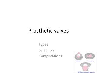
Valves Biologic (Tissue) Mechanical • Stented • Porcine xenograft • Pericardial xenograft • Stentless • Porcine xenograft • Pericardial xenograft • Homograft • Autograft • Ball and cage (Starr-Edwards) • Single tilting disc (Medtronic-Hall) • Bileaflet (St. Jude, CarboMedics)
Mechanical Valves • Extremely durable with overall survival rates of 94% at 10 years • Primary structural abnormalities are rare • Most malfunctions are secondary to perivalvular leak and thrombosis • Chronic anticoagulation required in all • With adequate anticoagulation, rate of thrombosis is 0.6% to 1.8% per patient-year for bileaflet valves
Biological Valves • Stented bioprostheses • Primary mechanical failure at 10 years is 15-20% • Preferred in patients over age 70 • Subject to progressive calcific degeneration & failure after 6-8 years • Stentless bioprostheses • Absence of stent & sewing cuff allow implantation of larger valve for given annular size->greater EOA • Uses the patient’s own aortic root as the stent, absorbing the stress induced during the cardiac cycle
Biologic Valves Continued • Homografts • Harvested from cadaveric human hearts • Advantages: resistance to infection, lack of need for anticoagulation, excellent hemodynamic profile (in smaller aortic root sizes) • More difficult surgical procedure limits its use • Autograft • Ross Procedure
Approach to Valve Evaluation • Clinical data including reason for the study and the patient’s symptoms • Type & size of replacement valve, date of surgery • BP & HR • HR particularly important in mitral and tricuspid evaluations because the mean gradient is dependent on the diastolic filling period • Patient’s height, weight, and BSA should be recorded to assess whether prosthesis-patient mismatch (PPM) is present
Echo Imaging of Prosthetic Valves • Valves should be imaged from multiple views, with attention to: • Opening & closing motion of the moving parts (leaflets for bioprosthesis and occluders for mechanical ones) • Presence of leaflet calcification or abnormal echo density attached to the sewing ring, occluder, leaflets, stents, or cage • Appearance of the sewing ring, including careful inspection for regions of separation from native annulus & for abnormal rocking motion during the cardiac cycle
Echo Imaging • Mild thickening is often the 1st sign of primary failure of a biologic valve • Occluder motion of a mechanical valve may not be well visualized by TTE because of artifact and reverberations
Imaging Considerations • Identify the sewing ring, valve or occluder mechanism, and surrounding area • Ball or disc is often indistinctly imaged, whereas leaflets of normal tissue valves should be thin with an unrestricted motion • Stentless or homograft may be indistinguishable from native valves • One can use modified views (lower parasternal) to keep the artifact from the valve away from the LV outflow tract
Doppler of Prosthetic AV • Doppler velocity recordings across normal PVs usually resemble those of mild native aortic stenosis • Maximal velocity usually > 2 m/s, with triangular shape of the velocity contour • Occurrence of maximal velocity in early systole • With increasing stenosis, a higher velocity and gradient are observed, with longer duration of ejection and more delayed peaking of the velocity during systole
Doppler Velocity Index (DVI) • Dimensionless ratio of the proximal velocity in the LVO tract to that of flow velocity through the prosthesis: • DVI= VLVO/ VPrAV • DVI is calculated as the ratio of respective VTIs and can be approximated as the ratio of respective peak velocities • Incorporates the effect of flow on velocity through the valve and is much less dependent on valve size
DVI • Helpful measure to screen for valve dysfunction, particularly when the CSA of the LVO tract cannot be obtained or valve size is unknown • DVI is always < 1 • DVI < 0.25 is highly suggestive of significant obstruction • DVI is not affected by high flow conditions through the valve, including AI
Doppler & Prosthetic AV • High gradients may be seen with normal functioning valves with: • Small size • Increased stroke volume • PPM • Valve obstruction • Conversely, a mildly elevated gradient in the setting of severe LV dysfunction may indicate significant stenosis • Thus, the ability to distinguish malfunctioning from normal PVs in high flow states on the basis of gradients alone may be difficult
Doppler Continued • Other qualitative and quantitative indices that are less dependent on flow should be evaluated • Contour of the velocity: • In a normal valve, even in high flow, there is a triangular shape, with early peaking of the velocity and short acceleration time (AT) • With PV obstruction, a more rounded velocity contour is seen, with velocity peaking almost in mid-ejection, prolonged AT • Cutoff of AT of 100 ms differentiates well between normal and stenotic PVs
Effective Orifice Area (EOA) • EOA PrAV = (CSA LVO x VTI LVO) / VTI PrAV • EOA is dependent on size of inserted valve • Should be referenced to the valve size of a particular valve type • For any size valves, significant stenosis is suspected when valve area is < 0.8 cm2 • However, for the smallest size valve, this may be normal because of pressure recovery • Largest source of variability is measurement of the LVO tract
Doppler Parameters of Prosthetic AV function in Mechanical and Stented Biologic Valves in Conditions of Normal Stroke Volume
Patient-Prosthesis Mismatch (PPM) • When the EOA of the inserted prosthesis is too small in relation to the patient’s BSA • A given valve area acceptable for a small, inactive person may be inadequate for a larger physically active individual • Main consequence is the generation of higher than expected gradients through a normally functioning valve
PPM Continued • Commonly seen in: • Patients with small aortic annulus sizes, particularly women • Patients whom indication for AVR was AS as opposed to AI • Young patients, who outgrow their initially inserted prosthesis • Failure of post-op regression of LV mass index at 6 months may be clue to presence of PPM • For patients with exertional symptoms without evidence of primary valve dysfunction, stress echo should be entertained to further evaluate
Evaluation of Prosthetic AI • With color doppler, one can evaluate the components of the color AI jet • Flow convergence, vena contracta, extent in the LVO tract and LV • Normal “physiologic” jet are usually low in momentum, depicted by homogenous color jets that are small in extent • Ratios of jet diameter/LVO diameter from parasternal long-axis imaging and Jet area/LVO area from parasternal short-axis imaging are best applied for central jets
Prosthetic Valve AI • With eccentric AI jets, measurement of jet width perpendicular to the LVO tract will cut the jet obliquely and risk overestimation • Entrainment of jet in the LVO tract may lead to rapid broadening of the jet just after the vena contracta-> overestimation
AI in PVs • Contrary to native valves, the width of the vena contracta may be difficult to accurately measure in the long-axis in the presence of a prosthesis • Imaging of the neck of the jet in short-axis, at the level of the sewing ring allows determination of the circumferential extent of the regurgitation • Approximate guide: • < 10% of sewing ring suggests mild • 10-20% suggests moderate • > 20% suggests severe • **Rocking of the prosthesis usually associated with >40% dehisscence
Spectral Doppler and PVAI • PHT is useful when the value is <200 ms, suggesting severe AI, or > 500 ms, consistent with mild AI • Intermediate ranges may reflect other hemodynamic variables such as LV compliance and are less specific • Holodiastolic flow reversal in the descending thoracic aorta is indicative of at least moderate AI • Severe is suspected when the VTI of the reverse flow approximates that of the forward flow • Holodiastolic flow reversal in the abdominal aorta is usually indicative of severe AI
Evaluation of Prosthetic MV • A major consideration with echo is the effect of acoustic shadowing by the prosthesis on assessment of MR • Problem is worse with mechanical valves • On TTE, LV function is readily evaluated, but the LA is often obscured for imaging and doppler interrogation • TEE provides visualization of the LA and MR but shadowing limits visualization of the LV • Thus, comprehensive assessment of PMV requires both TTE & TEE when valve dysfunction is suspected
Prosthetic MV Imaging Considerations • In the parasternal long-axis view, the prosthesis may obscure portions of the LA and its posterior wall • MR may be difficult to evaluate • Parasternal long-axis views allows visualization of the LVO tract, which can be impinged by higher profile prostheses • Apical views allow visualization of leaflet excursion for both bioprosthetic and mechanical valves • May allow detection of thrombus or pannus • Vegetations can be seen but are often masked by acoustic shadowing
Doppler Evaluation of PMV • Complete exam should include: • Peak early velocity • Estimate of mean pressure gradient • Heart Rate • Pressure half-time (PHT) • Determination of whether regurgitation is present • DVI and/or EOA as needed • LV/RV size and function • LA size if possible • PA systolic pressure
Peak Early Mitral Velocity • Peak E velocity is easy to measure • Provides simple screen for prosthetic valve dysfunction • Can be elevated in: hyperdynamic states, tachycardia, small valve size, stenosis, or regurgitation • Inhomogeneous flow profile across caged-ball and bileaflet prostheses can lead to doppler velocity measurements that are elevated out of proportion to the actual gradient • For normal bioprosthetic MVs, peak velocity can range from 1.0 to 2.7 m/s
MV Peak Velocity • In normal bileaflet mechanical valves, peak velocity is usually < 1.9 m/s but can be up to 2.4 m/s • As a general rule, peak velocity < 1.9 m/s is likely to be normal in most patients with mechanical valves unless there is markedly depressed LV function
Mean Gradients of MV • Normally less than 5-6 mm Hg • Values up to 10-12 mm Hg have been reported in normally functioning mechanical valves • High gradients can be due to: hyperdynamic states, tachycardia or PPM, regurgitation, or stenosis
MV Pressure Half-time (PHT) • A large rise in PHT on serial studies or a markedly prolonged single measurement (>200 ms) may be a clue to the presence of: obstruction • PHT seldom exceeds 130 ms across normal pv • Minor changes in PHT occur as a result of nonprosthetic factors including: • Loading conditions • Drugs • AI • PHT should not be obtained in tachycardic rhythms or 1st degree blocks when the E & A velocities are merged or the diastolic filling period is short
EOA of PMV • Calculation from PHT, as traditionally applied in native MS, is not valid in prosthetic valves due to its dependence on LV and LA compliance and initial LA pressure • EOAPrMV= stroke volume/VTIPrMV • Usually reserved for cases of discrepancy between information obtained from gradients and PHT
Prosthetic MV and DVI • DVI= VTIPrMV/ VTILVO • DVI can be elevated with stenosis or regurgitation • For mechanical valves, a DVI < 2.2 is most often normal • Higher values should prompt consideration of prosthesis dysfunction