Download
1 / 45
750 likes | 2.25k Views
CHIANGMAI UNIVERSITY. บทที่ 3. RBC MORPHOLOGY. Prathom Prathomthanapongs. CHIANGMAI UNIVERSITY. RBC MORPHOLOGY. การศึกษาลักษณะของเม็ดเลือดแดงเพื่อการวินิจฉัยภาวะของโรคต่างๆเกี่ยวกับการสร้างและการทำลายของเม็ดเลือดแดงนั้น ต้องทราบว่าการเปลี่ยนแปลงอาจเกิดได้จากหลายสาเหตุดังนี้ :-

E N D
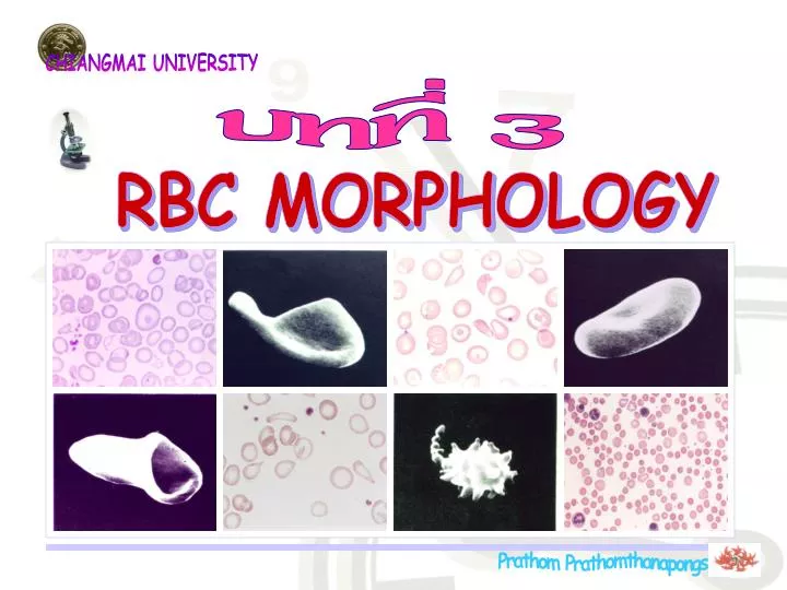
CHIANGMAI UNIVERSITY บทที่ 3 RBC MORPHOLOGY Prathom Prathomthanapongs
CHIANGMAI UNIVERSITY RBC MORPHOLOGY การศึกษาลักษณะของเม็ดเลือดแดงเพื่อการวินิจฉัยภาวะของโรคต่างๆเกี่ยวกับการสร้างและการทำลายของเม็ดเลือดแดงนั้นต้องทราบว่าการเปลี่ยนแปลงอาจเกิดได้จากหลายสาเหตุดังนี้:- 1.Abnormal erythropoiesis (Metabolic defects) 2.Acquired or induced in circulation 3.การพบเซลล์อ่อน Prathom Prathomthanapongs
CHIANGMAI UNIVERSITY Abnormal RBC Morphology การเปลี่ยนแปลงรูปร่าง ของเม็ดเลือดแดง จนทำให้ เกิดลักษณะที่ผิดปกติต่างๆนั้น มีการแบ่งการเปลี่ยนแปลงออก เป็นหลายลักษณะ ได้แก่:- 1. Anisocytosis (Abnormal in Size) 2. Poikilocytosis (Abnormal in Shape) 3. Color & Distribution 4. Red cell inclusions Prathom Prathomthanapongs
CHIANGMAI UNIVERSITY 1.Anisocytosis (Abnormal in size) 1.1 Discocyte (Normocyte) เม็ดเลือดแดงปกติมีบุ๋มกลางเรียกว่าbiconcave การติดสีจะจางที่กลางเซลล์ เรียก central pallor ขนาดของเซลล์คือ:- wet preparation 8.4 mm - dried blood film 7.2-7.4 mm - surface area 140 mm2 - ด้านข้าง -ส่วนหนา2.4 mm -ส่วนบุ๋มกลาง1 mm สามารถผ่านfilter ขนาด 3 mmได้ จะผ่านไม่ได้เมื่อมีปัญหาของการเกิด elastic deformation และ plastic formation Prathom Prathomthanapongs
CHIANGMAI UNIVERSITY 1.Anisocytosis (Abnormal in size) 1.2 Macrocyte เป็นเม็ดเลือดแดงที่มีขนาดใหญ่กว่า8 mm สาเหตุเกิดจาก:- - Abnormal erythropoiesis - Pernicious anemia ขาด Vit.B12,Folic acid - After acute blood loss - Hemolytic disease of newborn Prathom Prathomthanapongs
CHIANGMAI UNIVERSITY 1.Anisocytosis (Abnormal in size) 1.3 Microcyte เป็นเม็ดเลือดแดงที่มี ขนาดเล็กกว่า6.4 mm สาเหตุเกิดจาก:- - Iron deficiency - Inflammation Prathom Prathomthanapongs
CHIANGMAI UNIVERSITY 2.Poikilocytosis (Abnormal in shape) 2.1 Elliptocyte (Ovalocyte) เป็นเม็ดเลือดแดงที่มีลักษณะรูปรี ถ้าค่อนข้างกลมคล้ายไข่ไก่เรียกOvalocyteถ้ายาวเรียกElliptocyte ปกติพบ< 1% ถ้าพบมากกว่าปกติ เกิดจากสาเหตุดังนี้:- - พบ < 10 % - Megaloblastic anemia - Iron deficiency - Myelofibrosis - พบ> 50 % - Hereditary elliptocytosis Prathom Prathomthanapongs
CHIANGMAI UNIVERSITY 2.Poikilocytosis (Abnormal in shape) 2.2 Spherocyte เป็นเม็ดเลือดแดงที่มีลักษณะกลมไม่เป็น biconcave ส่วนใหญ่จะมีขนาดเล็กกว่าปกติเรียกmicrospherocyte เม็ดเลือดแดงพวกนี้มักจะมีอายุสั้น พื้นที่ผิวลดลงและมีผนัง เซลล์หนามากกว่าปกติ - พบได้เล็กน้อยในภาวะ:- - Hemolytic mechanism จากสาเหตุ chemical & bacterial toxins - พบจำนวนมากใน:- Hereditary spherocytosis Prathom Prathomthanapongs
CHIANGMAI UNIVERSITY 2.Poikilocytosis (Abnormal in shape) 2.3 Dacryocyte (Teardrop Cell) เป็นเม็ดเลือดแดงที่มีลักษณะเป็นรูปหยดน้ำตา ไม่พบใน คนปกติ เกิดจากสาเหตุ “Pitting” เม็ดเลือดแดงที่ผิดปกติของ R.E. system - พบได้ในภาวะ:- - Thalassemia - Drug induced “Heinz bodies” - Extramedullary erythropoiesis - Myeloid metaplasia “ Myelofibrosis ” Prathom Prathomthanapongs
CHIANGMAI UNIVERSITY 2.Poikilocytosis (Abnormal in shape) 2.4 Drepanocyte (Sickle cell) - เป็นเม็ดเลือดแดงที่มีลักษณะเป็นรูปเคียว หรือเรียกว่า“Boat shape” - มีพื้นที่ผิวมากกว่าปกติ - มี mechanical fragility สูง - พบได้ใน”Hb S disease” Prathom Prathomthanapongs
CHIANGMAI UNIVERSITY 2.Poikilocytosis (Abnormal in shape) 2.5 Codocyte (Target cell) เป็นเม็ดเลือดแดงที่มีลักษณะเป็นรูปเป้า มีลักษณะเป็น “Cup shape” ใน wet preparation มีพื้นที่ผิวมากกว่าปกติ - พบได้ในภาวะ:- - Obstructive liver disease - Deficiency of enzyme LCAT “Lecithin-choresterol acyl transferase” - Iron deficiency anemia พบน้อย - พบมากใน Thalassemia Prathom Prathomthanapongs
CHIANGMAI UNIVERSITY 2.Poikilocytosis (Abnormal in shape) 2.6 Stomatocyte เป็นเม็ดเลือดแดงที่มีลักษณะเป็น “Cup shape” ใน wet preparation คล้าย Codocyte/Target cell) - พบได้ในภาวะ:- - Cationic drugs “ pH “ - Liver disease - Alcohol intake - พบมากใน Hereditary stomatocytosis Prathom Prathomthanapongs
CHIANGMAI UNIVERSITY 2.Poikilocytosis (Abnormal in shape) 2.7 Echinocyte เป็นเม็ดเลือดแดงที่มีลักษณะเป็นเซลล์เหี่ยว มีส่วนยื่นมากและมีความยาวเท่าๆกัน พบปกติได้เล็กน้อย พบมากในภาวะที่มี“ pH “ Prathom Prathomthanapongs
CHIANGMAI UNIVERSITY 2.Poikilocytosis (Abnormal in shape) 2.8 Acanthocyte ( Spur cell ) เป็นเม็ดเลือดแดงที่พบส่วนยื่น 3-20 อัน มีความยาวไม่เท่ากัน ดูด้วยกล้อง EMปลายมน ส่วนกล้องจุลทรรศน์ธรรมดาปลายจะแหลม - พบได้เล็กน้อยใน:- - Alcoholic liver cirrhosis - Hepatitis - พบสูงใน:- - Hereditary abetalipoproteinemia Prathom Prathomthanapongs
CHIANGMAI UNIVERSITY 2.Poikilocytosis (Abnormal in shape) 2.9 Keratocyte เป็นเม็ดเลือดแดงที่พบส่วนยื่นประมาณ 2 อัน เกิดจากแรงกระแทกของเส้น fibrin หรือลิ้นหัวใจเทียม - และพบได้ใน:- - Uremia - Glomerulonephritis - DIC (Disseminated intravascular coagulation) Prathom Prathomthanapongs
CHIANGMAI UNIVERSITY 2.Poikilocytosis (Abnormal in shape) 2.10 Schistocyte ( Fragmented cell ) ลักษณะเป็นชิ้นส่วนของเม็ดเลือดแดงเกิดจาก:- - Physical/Mechanical stress ต่อเม็ดเลือดแดง เช่น:- - Drugs - Chemical - Hemoglobin instability in thalassemia - DIC (Disseminated intravascular coagulation) Prathom Prathomthanapongs
CHIANGMAI UNIVERSITY 3. Color & Distribution 3.1 Color Polyochromasia Normochromia การย้อมติดสีของเม็ดเลือดแดง ขึ้นกับปริมาณของ hemoglobin ที่มีในเซลล์ แบ่งชนิดการติดสีได้ดังนี้:- - Normochromia - Hypochromia - Polychromasia - Hyperchromia ในบางรายสามารถพบเม็ดเลือดแดง ติดสีแตกต่างกันมากกว่าหนึ่งชนิดเรียกว่า “Dimorphism” “Dimorphism” Hypochromia Prathom Prathomthanapongs
CHIANGMAI UNIVERSITY 3. Color & Distribution Dimorphism มีเม็ดเลือดแดงย้อมติดสีแบบ “Normochromia หรือ Hypochromia” อยู่ปะปนปนกัน สามารถพบได้ในภาวะ ต่อไปนี้:- - Iron deficiency- after treatment - After blood transfusion - Sideroblastic anemia (Hereditary or acquired) - Megaloblastic anemia Prathom Prathomthanapongs
CHIANGMAI UNIVERSITY 3. Color & Distribution 3.2 Distribution Rouleaux Formation ลักษณะเรียงซ้อนกันของเม็ดเลือดแดง สามารถพบได้ในภาวะต่อไปนี้:- - Hyperproteinemia - Macroglobulinemia - Multiple myeloma Prathom Prathomthanapongs
CHIANGMAI UNIVERSITY 3. Color & Distribution 3.2 Distribution Agglutination ลักษณะการจับกลุ่มกันของเม็ดเลือดแดง สามารถพบได้ในภาวะต่อไปนี้:- - ปฏิกิริยา Antigen - Antibody - Cold hemagglutination - Paroxysmal cold hemoglobinuria Prathom Prathomthanapongs
CHIANGMAI UNIVERSITY 4. Red Cell Inclusions 4.1 Howell-Jolly Bodies เป็นก้อนDNAขนาด 1-2 mm สีม่วง-น้ำเงินเข้มสามารถพบได้ในภาวะ ต่อไปนี้:- - Megaloblastic anemia - Thalassemia - Splenectomy Prathom Prathomthanapongs
CHIANGMAI UNIVERSITY 4. Red Cell Inclusions 4.2 Basophilic Stippling เป็นลักษณะจุดเล็กๆในเม็ดเลือดแดง ปกติไม่พบในผู้ใหญ่ อาจพบได้ในเด็กอ่อน หรือเด็กเล็กและสามารถพบได้ในภาวะ ต่อไปนี้:- - Lead poisoning - Pyrimidine-5-nucleotide def. - Disorders of Hb synthesis เรียก ”Punctate basophilia” พบใน”Homozygous b thalassemia” Prathom Prathomthanapongs
CHIANGMAI UNIVERSITY 4. Red Cell Inclusions 4.3 Cabot's Ring เป็นส่วนหลงเหลือของ ”Spindle fibers” ในขณะเซลล์แบ่งตัว สามารถพบได้ในภาวะต่อไปนี้:- - Severe anemia - Dyserythropoiesis - Splenectomy Prathom Prathomthanapongs
CHIANGMAI UNIVERSITY 4. Red Cell Inclusions 4.4 Heinz Bodies มีลักษณะเป็นก้อนกลมขนาด0.3-3.0 mmของฮีโมโกลบินที่แปลงสภาพย้อมด้วยสี ”Supravital stain” สามารถพบได้ในภาวะต่อไปนี้:- - G-6-PD deficiency - Thalassemia - Unstable hemoglobin เช่นHb H(b4) inclusion bodies - ได้รับสาร oxidant Prathom Prathomthanapongs
CHIANGMAI UNIVERSITY 4. Red Cell Inclusions 4.5 Malaria เป็นเม็ดเลือดแดง ที่มีการติดเชื้อ malaria ลักษณะแล้วแต่ชนิดและระยะ ของเชื้อmalariaนั้นๆ Prathom Prathomthanapongs
CHIANGMAI UNIVERSITY 4. Red Cell Inclusions 4.6 Pappenheimer Bodies เป็นgranules ของเหล็กใน เม็ดเลือดแดงที่เกิดจากการรวม ตัวของเหล็กกับmitochondria และ ribosomes สามารถพบได้ใน:- - Severe anemias - Thalassemias. Prathom Prathomthanapongs
CHIANGMAI UNIVERSITY 1 Grading of RBC Morphology การรายงานลักษณะรูปร่างของเม็ดเลือดแดงด้วยกล้องจุลทรรศน์ เพื่อใช้วินิจฉัยโรคเลือดที่เกี่ยวกับการสร้างและการทำลายของเม็ดเลือดแดงนั้นต้องพิจารณาถึงรายละเอียดต่างๆดังต่อไปนี้:- 1. เป็นสเมียร์เลือด จากเลือดที่ใหม่ ไม่ค้างอยู่ ในสารกันเลือดแข็งนานเกินไป หรือเป็นเลือด ที่มีอัตราส่วนของสารกันเลือดแข็งต่อเลือด มากเกินไปเพราะจะทำให้รูปร่างของเม็ดเลือด แดงเปลี่ยนไป 2. เป็นสเมียร์จากเลือดที่แห้งสนิทก่อนย้อมสี (air-dried) Prathom Prathomthanapongs
CHIANGMAI UNIVERSITY 2 Grading of RBC Morphology 3. การรายงานรูปร่างเม็ดเลือดแดงให้รายงานจาก การตรวจสเมียร์เลือดบริเวณ ideal area เท่านั้น เพราะจะเป็นบริเวณที่เม็ดเลือดแดงเรียงตัวไม่ซ้อนกัน และไม่ห่างกันมากเกินไปโดยตรวจจำนวนไม่น้อยกว่า 10 oil fields ซึ่งจะมีจำนวนเม็ดเลือดแดง ประมาณ 200 เซลล์ต่อ 1 oil field ในคนปกติ ideal area Prathom Prathomthanapongs
CHIANGMAI UNIVERSITY 3 Grading of RBC Morphology ิ 4. เนื่องจากเม็ดเลือดแดงมีอายุในกระแสเลือดประมาณ 120 วัน ทำให้คนปกติอาจมีเม็ดเลือดแดงที่มีรูปร่างและขนาดผิดปกติบ้าง ตาม ความแตกต่างของอายุเม็ดเลือดแดง ดังนั้นการรายงานความผิดปกติ ของเม็ดเลือดแดง จะต้องรายงานเมื่อมีความผิดปกติมากกว่าจำนวน ที่พบได้ในคนปกติ สำหรับเม็ดเลือดแดงผิดปกติที่ไม่พบในคนปกติต้อง รายงานทุกครั้งที่พบไม่ว่าจะพบมากหรือน้อยก็ตาม ลักษณะ ความผิดปกติของเม็ดเลือดแดงมีหมวดใหญ่ๆอยู่ 4 หมวด คือ:- - Anisocytosis(Abnormal in Size) - Poikilocytosis(Abnormal in Shape) - Color & Distribution - Red cell inclusions Prathom Prathomthanapongs
CHIANGMAI UNIVERSITY 4 Grading of RBC Morphology เกณฑ์การให้คะแนนระดับความผิดปกติในปัจจุบัน นิยมให้เป็น Grading:- few ,1+,2+,3+ &4+ โดยรายงานเป็นระดับตามปริมาณที่พบดังนี้:- รายงานfew =-ทุกครั้งที่พบและไม่เกิน 10 % สำหรับเซลล์ที่ไม่พบในคนปกติได้แก่:- - macroovalocyte,acanthocyte, -teardrop cell,spherocyte ( รวมทั้ง microspherocyte) ถ้าพบจะต้องรายงาน รายงาน1+=พบเซลล์ผิดปกติจำนวน 11-25 % รายงาน2+ = พบเซลล์ผิดปกติจำนวน 26-50 % รายงาน3+ = พบเซลล์ผิดปกติจำนวน 51-75 % รายงาน4+= พบเซลล์ผิดปกติจำนวน >75 % Prathom Prathomthanapongs
CHIANGMAI UNIVERSITY 5 Grading of RBC Morphology จากหมวดและเกณฑ์การรายงานความผิดปกติของรูปร่างเม็ดเลือดแดง สามารถรายงานผลได้ดังนี้:- 4.1 Anisocytosis(Abnormal in Size) การรายงานความผิดปกติของขนาดเม็ดเลือดแดงให้ รายงานในภาพรวมว่ามีความผิดปกติของขนาดว่าเล็กเกินไป หรือใหญ่เกินไปมีปริมาณเท่าไร (few ,1+,2+,3+ & 4+) แล้งจึงรายงานแยกชนิดว่ามีขนาดเล็กหรือขนาดใหญ่อย่าง ละเท่าไร (few ,1+,2+,3+ & 4+) ซึ่งผลรวมไม่จำเป็นว่า จะต้องเท่ากับผลของภาพรวม แต่ไม่ควรจะมากกว่า Prathom Prathomthanapongs
CHIANGMAI UNIVERSITY 6 Grading of RBC Morphology 4.2 Poikilocytosis(Abnormal in Shape) การรายงานความผิดปกติรูปร่างของเม็ดเลือดแดงให้รายงาน ในภาพรวมว่ามีความผิดปกติของรูปร่างว่า มีปริมาณเท่าไร (few ,1+,2+,3+ & 4+)แล้วจึงรายงานแยกทุกชนิดที่พบว่ามี รูปร่างอะไรบ้างที่มีความผิดปกติ ( โดยต้องเป็นจำนวนที่มากกว่าปกติที่ควรมี)ว่ามีปริมาณเท่าไร (few ,1+,2+,3+ &4+) ซึ่งผลรวมไม่จำเป็นว่าจะต้องเท่ากับผลของภาพรวม หมายเหตุ ในกรณีที่พบเซลล์มีความผิดปกติทั้งขนาดและรูปร่างในเซลล์เดียวกัน เช่น microspherocyte หรือ macroovalacyte ให้รายงายแยกชนิดในแต่ละช่องของรายงาน และสามารถรายงาน ให้เห็นชัดเจนขึ้นในช่องของ Others & Immpression ว่ามี microspherocyte หรือ macroovalacyte ปริมาณเท่าไรด้วย (few ,1+,2+,3+ &4+) Prathom Prathomthanapongs
CHIANGMAI UNIVERSITY 7 Grading RBC morphology 4.3 Color & Distribution 4.3.1 Hypochromia ให้รายงานความผิดปกติของปริมาณ hemoglobin ในเม็ดเลือดแดงที่พบเกิน2 % โดยให้นับจำนวน เซลล์ที่มี central pallorมากกว่า 1/3 โดยรายงานเป็น few ,1+,2+,3+ & 4+ตามจำนวนที่พบ 4.3.2 Polychromasiaให้รายงานทุกครั้งที่พบโดยนับจำนวน ใน 10 oil fields แล้วเฉลี่ยตามเกณฑ์ดังนี้:- รายงานfewเมื่อพบ polychromasia 0-1 cells/OF รายงาน1+ เมื่อพบ polychromasia 2-3 cells/OF รายงาน 2+ เมื่อพบ polychromasia 4-6 cells/OF รายงาน3+ เมื่อพบ polychromasia 7-12 cells/OF รายงาน 4+ เมื่อพบ polychromasia >12 cells/OF Prathom Prathomthanapongs
CHIANGMAI UNIVERSITY 8 Grading RBC morphology 4.3.3 Distribution ให้รายงานความผิดปกติของ การเรียงตัวของเม็ดเลือดแดงโดยรายงานทุกครั้งที่พบ เช่น:- การพบ rouleaux formation เป็นต้น 4.4 Red cell inclusions ให้รายงานทุกครั้งที่พบโดย รายงานแต่เพียงว่า “ Presentหรือ foundหรือ พบ ” โดยไม่ต้องแบ่ง ระดับการพบ Red cell inclusions ทั่วๆไป ยกเว้นmalaria น่าจะนับจำนวนเชื้อ malaria ที่พบ ต่อเม็ดเลือดแดงจำนวน 1,000 หรือ 10,000 เซลล์ ตามจำนวนของเชื้อ เพื่อเป็นการให้เห็นภาพของภาวะ การติดเชื้อ malaria ในเลือดของผู้ป่วยได้ชัดเจนมากขึ้น Prathom Prathomthanapongs
CHIANGMAI UNIVERSITY Grading of RBC Morphology Slide no.1 Normal (normochromic normocytic) Abnormal RBC morphology Anisocytosis few Microcyte Macrocyte Poikilocytosis 1+ Target cell few Spherocyte Fragmented cell few Hypochromia 1+ Polychromasia RBC inclusion Thalassemia carrier OTHERS & IMPRESSION: Prathom Prathomthanapongs
CHIANGMAI UNIVERSITY Grading of RBC Morphology Slide no.2 Normal (normochromic normocytic) Abnormal RBC morphology Anisocytosis few Microcyte Macrocyte Poikilocytosis 1+ Target cell few Spherocyte Fragmented cell few Tear drop few Hypochromia 1+ Polychromasia RBC inclusion Thalassemia carrier OTHERS & IMPRESSION: Prathom Prathomthanapongs
CHIANGMAI UNIVERSITY Grading of RBC Morphology Slide no.3 Normal (normochromic normocytic) Abnormal RBC morphology Anisocytosis 2+ Microcyte 1+ Macrocyte 1+ Poikilocytosis 3+ Target cell 1+ Spherocyte Fragmented cell 1+ Hypochromia 4+ Polychromasia Howell jolly body Present Thalassemia diseases OTHERS & IMPRESSION: Prathom Prathomthanapongs
CHIANGMAI UNIVERSITY Grading of RBC Morphology Slide no.4 Normal (normochromic normocytic) Abnormal RBC morphology Anisocytosis 1+ Microcyte Macrocyte Poikilocytosis 3+ Target cell 1+ Spherocyte Fragmented cell 1+ Hypochromia 3+ Polychromasia RBC inclusion NRBC Present Thalassemia diseases OTHERS & IMPRESSION: Prathom Prathomthanapongs
CHIANGMAI UNIVERSITY Grading of RBC Morphology Slide no.5 Normal (normochromic normocytic) Abnormal RBC morphology Anisocytosis 2+ Microcyte 1+ Macrocyte few Poikilocytosis 3+ Target cell few Spherocyte 1+ Fragmented cell 2+ Hypochromia 2+ Polychromasia RBC inclusion Thalassemia diseases OTHERS & IMPRESSION: Prathom Prathomthanapongs
CHIANGMAI UNIVERSITY Grading of RBC Morphology Slide no.6 Normal (normochromic normocytic) Abnormal RBC morphology Anisocytosis 3+ Microcyte 3+ Macrocyte few Poikilocytosis 3+ Target cell Spherocyte Fragmented cell 1+ Tear drop few Hypochromia 4+ Polychromasia RBC inclusion OTHERS & IMPRESSION: Iron deficiency anemia Prathom Prathomthanapongs
CHIANGMAI UNIVERSITY Grading of RBC Morphology Slide no.7 Normal (normochromic normocytic) Abnormal RBC morphology Anisocytosis 2+ Microcyte few Macrocyte 2+ Poikilocytosis 1+ Target cell Spherocyte Fragmented cell few Hypochromia 2+ Polychromasia RBC inclusion OTHERS & IMPRESSION: Megaloblastic anemia Prathom Prathomthanapongs
CHIANGMAI UNIVERSITY Grading of RBC Morphology Slide no.8 Normal (normochromic normocytic) Abnormal RBC morphology Anisocytosis few Microcyte few Macrocyte Poikilocytosis 1+ Target cell Spherocyte Fragmented cell few Hypochromia 2+ Polychromasia RBC inclusion OTHERS & IMPRESSION: Dimorphism Prathom Prathomthanapongs
CHIANGMAI UNIVERSITY Grading of RBC Morphology Slide no.9 Normal (normochromic normocytic) Abnormal RBC morphology Anisocytosis 2+ Microcyte 1+ Macrocyte few Poikilocytosis 2+ Target cell Spherocyte Fragmented cell few Tear drop 1+ Hypochromia few Polychromasia RBC inclusion Tear drop cells OTHERS & IMPRESSION: Prathom Prathomthanapongs
CHIANGMAI UNIVERSITY Grading of RBC Morphology Slide no.10 Normal (normochromic normocytic) Abnormal RBC morphology Anisocytosis 4+ Microcyte 3+ Macrocyte few Poikilocytosis 3+ Target cell Spherocyte 3+ Fragmented cell Hypochromia Polychromasia RBC inclusion NRBC Present OTHERS & IMPRESSION: Microspherocytosis Prathom Prathomthanapongs
BLOOD & BONE MARROW CELLS สวัสดี CHIANGMAI UNIVERSITY