Download
1 / 24
240 likes | 536 Views
Chlamydiae. By. Dr. Emad AbdElhameed Morad. Lecturer of Medical Microbiology and Immunology. Chlamydiae were classified in the past as large viruses because they are: Small in size (250 – 400 nm). Obligatory intracellular because they can not make their own ATP.

E N D
Chlamydiae By Dr. Emad AbdElhameed Morad Lecturer of Medical Microbiology and Immunology
Chlamydiae were classified in the past as large virusesbecause they are: • Small in size (250 – 400 nm). • Obligatory intracellular because they can not make their own ATP. • Form intracytoplasmic inclusion bodies inside the host cells.
However, chlamydiae differ from virusesand simulate bacteriain the following: • They have cell wallsimilar to Gram negative bacteria. • They have both DNA and RNA. • They contain plasmids. • They have ribosomes and synthesize their own proteins. • They have metabolically active enzymes. • They are sensitive to antibiotics. • They replicate by a special cycle including binaryfission.
Developmental cycle It begins when the elementary body (300 nm)is taken inside the cell by phagocytosis like process. Inside the cell, the elementary body becomes initial or reticulate body. The reticulate bodygrows in size and divide by binary fission forming large number of elementary bodies. These elementary bodies are seen inside the cells as intracytoplasmic inclusion bodies. Elementary bodiesare then released to infect new cells.
Morphology & Cultural characters Chlamydia is cultivated on: Tissue culture (McCoy cells or HEP-2 cells) Chick embryo Inside the cells, chlamydia form intracytoplasmic inclusion bodies which could be stained by Giemsa or Macchiavello stains.
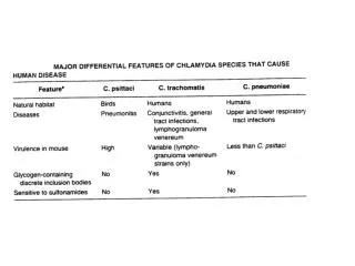
Diseases • Serotypes A, B, C cause: • Ocular infection called trachoma. • Trachoma is transmitted from eye to eye by fingers, flies and fomites.
Serotypes D-K cause: • Genital infection: • Transmitted sexually. • In males:non gonococcal urethritis. • In females:cervicitis and salpingitis. • Ocular infection: • Inclusion conjunctivitis. • Respiratory infections: • Upper respiratory infections like pharyngitis and otitis media. • Pneumonia may occur in immunocompromised patients.
Non gonococcal urethritis Cervicitis Salpingitis Inclusion conjunctivitis
Reiter’s syndrome? • Patients with genital chlamydia trachomatis infectionmay develop Reiter’s syndrome. • Reiter’s syndrome= urethritis + arthritis + uveitis. • This occurs because the antibodies produced against Chlamydia trachomatiscross-react with the antigens on urethra, joints and uveal tract.
Serotypes L1, L2, L3 cause: • Lymphogranuloma venereum (LGV): • Sexually transmitted disease. • Frie test is a skin test similar to tuberculin test and could be used for diagnosis.
Disease • Chlamydophila psittacicauses psittacosis. • Man gets infected by inhalation of dust containing dried faeces of birdsparticularly parrots. • The patient suffers from atypical pneumonia.
Disease • Chlamydophila pneumoniaecauses: Atypical pneumonia In humans only
Specimen: According to the site of infectionlike urethral discharge, sputum. Direct microscopic examination: After staining using Giemsa stain or immunofluorescent stain. To demonstrate intracytoplasmic inclusion bodies. Culture: Tissue culture: McCoy cellsfor Chlamydia trachomatis and Chlamydophila psittaci. HEP-2 cellsfor Chlamydophila pneumonia. Inclusion bodieswill be detected in tissue culture cells.
Direct detection in clinical specimen: For detection of the antigen: ELISA & IF. For detection of nucleic acid: PCR & DNA probes. Serological diagnosis: Detection of IgM or rising titer of IgGby ELISA or CFT.
Treatment • Macrolides like erythromycin and azithromycin. • Tetracyclines like doxycycline. • Patients with gonorrhea should also be treated for Chlamydia trachomatis.