Download
1 / 41
1.21k likes | 3.74k Views
Bone and alveolar bone. Periodontium (cont.). Dr Jamal Naim PhD in Orthodontics. Functions of bone. Support and protect the skeletal functions. Bone stores minerals, especially calcium and phosphorous, which are mobilized according to the body needs. Bone protects the internal organs.

E N D
Bone and alveolar bone Periodontium (cont.) Dr Jamal Naim PhD in Orthodontics
Functions of bone • Support and protect the skeletal functions. • Bone stores minerals, especially calcium and phosphorous, which are mobilized according to the body needs. • Bone protects the internal organs. • Bone marrow manufactures the blood elements. • Bone remodeling is responsible for the development, growth, movements, fracture and repair.
Osteoprogenitor cell Location: Present in the deepest layer of periosteum and lining the vascular canals of compact bone. Origin: It is mesenchymal in origin. Function: According to the function needed they divide by mitosis to give any type of bone cells Morphology: It resembles mesenchymal cell with pale stained nucleus and little esinophilic cytoplasm.
Bone cells All types of bone cells are from the same type, the alters there appearance according to there function: this is what is called cell modulation.
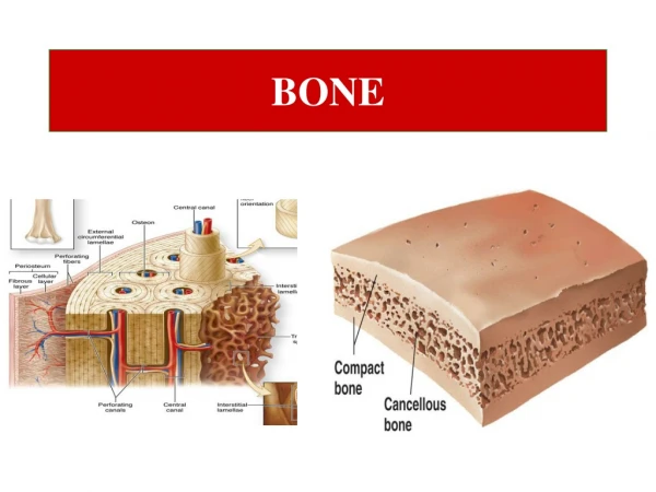
Types of bone Compact bone Spongy bone
Lamellar bone • Compact bone (ivory bone): It forms the main part of the shafts of the long bone and covers the cancellous bone e.g. ribs and flat bones of the skull. Its lamellae are arranged in 3 patterns: • Circumferential lamellae • Havarsian lamellae • Interstitial lamellae
Lamellar bone Circumferential lamellae: • outer circumferential lamellae beneath the periosteum. • inner circumferential lamellae adjacent to the endostium
Lamellar bone Havarsian lamellae: • It is the unite structure of the compact bone and is called the Haversian system or osteon. • The Haversian system if formed of Haversian canal which is surrounded by (4-20) concentrically arranged lamellae. • The Haversian canal contains B.Vs., nerves, C.T. and lined by osteoprogenitor cells.
Lamellar bone • Osteocytes are arranged concentrically in the osteons (they anastomose with each others by their process). • Haversian canals connected to each others or to the outer surface or to the bone marrow spaces with Volkmann's canal.
Lamellar bone Interstitial lamellae: • The Haversian systems are separated from each others by the interstitial lamellae. • They represent the remnants of an old resorbed and remodeled bone.
Bone Trabeculae Bone Marrow Spaces
Spongy bone • It is present in the central part of the flat bone. • It is formed of connected bone trabeculae in the form of network to give maximum rigidity. • In between the bone trabeculae, there are bone marrow spaces • The bone trabeculae have osteocytes.
Woven bone This type of bone is characterized by: • Irregular arrangement of the collagen fibers. • Great number, large size and irregular arrangement of the osteocytes. • Increase in the organic substance and decrease in the inorganic contents so; it appears radiolucent in X-ray. • This type of bone is resorbed completely and is replaced by lamellar bone.
3. Bundle Bone It is referred to the bundles of principal fibers of either the periosteum or PDL continue into the bone as sharpeys fibers.
Bundle bone • It is found adjacent to the periosteum and periodontal ligament (areas of tension). • It is characterized by the presence of the Sharpey's fibers. • It has less number of cells than Woven bone; but more calcium salts than lamellar bone. So it appears more radio-opaque and called lamina dura. • Its fibers are arranged parallel to the socket wall.
Alveolar bone In X-ray the cribriform plate is referred to lamina dura.
Alveolar bone The alveolar process is that bone containing the alveoli. It consists of: • an outer (lingual and buccal) cortical plate (compact bone) • A central spongiosa (spongous bone) and • Alveolar bone (bone lining the alveolus), (bundle bone) The alveolar bone and the cortical plate meet at the alveolar crest (1.5 to 2 mm below the level of CEJ).
Alveolar bone divided into the: • alveolar bone proper • lining of the tooth socket or alveolus • bone is also called the cribriform plate because of the many holes through which Volkmann’s canals pass (from the alveolar bone into the PDL)
Alveolar bone • also called bundle bone because Sharpey’s fibers insert into this bone (Sharpey’s fibers = portion of the fibers of the PDL) • these fibers are inserted at a 90 angle into the ABP – but are fewer in number than those found at the cemental surface • consists of plates of compact bone that surround the tooth
Alveolar bone • varies in thickness from 0.1 to 0.5mm • can see a portion of the ABP on radiographs lamina dura • most cervical rim = alveolar crest – slightly apical to the CEJ in healthy patients
Alveolar bone b. supporting alveolar bone • has the same components as ABP • but is considered to be cortical and trabecular bone – different arrangement of bony plates • cortical bone is made up of cortical plates of compact bone found on the facial and lingual surfaces • plates are usually 1.5 to 3mm thick over the posterior teeth and can vary over the anterior teeth
Alveolar bone • trabecular bone is located between the ABP and the plates of the cortical bone (cross section of mandible)
Alveolar bone • alveolar bone can be resorbed with age (edentulous) • the underlying basal bone is less affected with age – because it does not need the presence of teeth to remain viable • loss of teeth + alveolar bone can results in loss in the vertical dimension of the face – “Popeye” facial appearance
Alveolar bone • after tooth extraction the clot is replaced with immature bone • later remodelled as mature secondary bone, very similar process to fracture repair in skeletal bone
alveolar crest Cortical plate alveolar bone
Alveolar bone The alveolar bone is perforated by many foramina to allow blood and nerve supply to the teeth, so it is referred to cribriform plate Hirschfeld canal
Alveolar bone Collagen fibers
Alveolar bone Outer compact bone Spongiosa Collagen fibers
Alveolar bone Cementum Spongiosa Dentin PDL