Download
1 / 56
560 likes | 568 Views
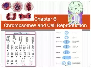
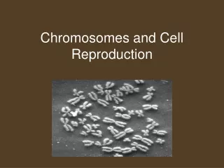
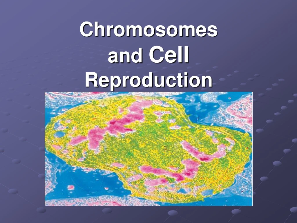
Chromosomes and Cell Reproduction. Chromosome Structure. Chromosomes are the coiled up version of DNA. They consist of DNA and proteins: histones and nonhistones. Chromosome Packing Animation. sister chromatids. centromere. single-stranded chromosomes. double-stranded chromosomes.

E N D
Chromosome Structure • Chromosomes are the coiled up version of DNA. • They consist of DNA and proteins: histones and nonhistones Chromosome Packing Animation
sister chromatids centromere single-stranded chromosomes double-stranded chromosomes • Point of Confusion: Chromatin vs. Chromatid vs. Chromosome • What is the Difference???
Chromatids are attached at the center called a centromere. Sister Chromatids
What is a Kinetochore? Kinetochore Animation
Centrosome & Centrioles oh the definitions… • Centrioles: ANIMAL CELLS ONLYNonmembrane-bound organelles that occur in pairs just outside the nucleus of animal cells. Each centriole is composed of a cylinder or ring of 9 sets of microtubule triplets with none in the middle (9 + 0 pattern). During cell division a pair of centrioles moves to each end of the cell, forming the poles of the mitotic spindle. Centrioles also give rise to basal bodies that control the origin of cilia and flagella in motile cells of protists. In cross section, flagella and cilia have 9 sets of microtubule doublets surrounding a pair of single microtubules in the center (9 + 2 pattern). This characteristic pattern also occurs in motile cells of higher organisms, such as human sperm. • Centrosome:In animal cells the centrosome includes a pair of centrioles surrounded by radiating strands of microtubules called the aster. The microtubule organizing centre that forms the mitotic spindle in dividing cells.
In Plants there is no Centriole • They use MTOCs: Microtuble Organizing Centers • Plant cells create mitotic spindle fibers and have a centrosome they lack centrioles.
What are spindle fibers made of? • Microtubles • Microtubles grow from the centrosomes • Polar Microtubles: Attach to other polar microtubles spaning the cell. • Kinetochore microtubles: attach to the kinetochore. Breakdown From Here. Microtubule formation and disassembly
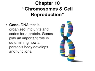
A gene is a segment of DNA that codes for a protein or RNA molecule which guide the development of traits.
Cell Types • Somatic Cells • Gametes
Chromosome Numbers • Types: Autosomes and Sex Chromosomes (XX or XY). • Humans: 22 pair of homologous chromosomes and 1 pair of sex chromosomes.
Chromosome Numbers • All of the cells in the body, other than gametes are Diploid (2n=46 in humans). • Gametes contain only one set of chromosomes: Haploid (n=23 in humans).
Change in chromosome number • Humans who are missing even one of the 46 chromosomes usually do not survive. • Humans with more than two copies of a chromosome, result in a condition called trisomy.
Abnormalities in chromosome number can be detected by analyzing a karyotype. • Nondisjunction during the production of gametes results in gametes having more or less chromosomes than normal. • Pictures called karyotypes can detect problems after conception.
How do Prokaryotes divide? • They divide by binary fission. Their DNA is circular and usually only has one strand. • No Mitosis!!!
Bacterial cells divide to reproduce • Binary fission is a form of asexual reproduction that produces identical offspring: Mitosis
The Cell Cycle: for EukaryotesFig 12.5,12.4, 12.17 • The cell cycle is a repeating sequence of cellular growth and division during the life of an organism. • A cell spends 90 percent of its time in the first three phases of the cycle.
I’m working here! Time to divide& multiply!
Key checkpoints at which feedback signals from the cell can trigger the next phase of the cell cycle (green light). Overview of the cell cycle Cell Cycle Movie/ Game
Checkpoint control system • 3 major checkpoints: • G1/S: Called “Restriction Point” • can DNA synthesis begin? • G2/M • has DNA synthesis been completed correctly? • commitment to mitosis • spindle checkpoint • are all chromosomes attached to spindle? • can sister chromatids separate correctly?
G1/DNA Synthesis checkpoint • G1/S checkpoint is most critical • primary decision point • “restriction point” • if cell receives “GO” signal, it divides • internal signals: cell growth (size), cell nutrition • external signals: “growth factors” • if cell does not receive signal, it exits cycle & switches to G0 phase • non-dividing, working state
G0 phase • G0 phase • non-dividing, differentiated state • most human cells in G0 phase • liver cells • in G0, but can be “called back” to cell cycle by external cues • nerve & muscle cells • highly specialized • arrested in G0 & can never divide
What molecules control cell division? • 2 types of molecules control • Cyclins oscillating levels during cell division. • Cycline dependent kinasis or CDKs • First CDK discovered was MPF/ p-phase promotor factor.
When control is lost: cancerFig 12.19, 12.20 • Cancer is the uncontrolled growth of cells due to loss of contact inhibition / density-dependent inhibition. • Overcrowding and tumors • Mutations in genes that control cell division may result in cancer. • If Telomerase ends are left on DNA, cells don’t die after 50 or so divisions. (Built in destruction control) • HeLa Cells: Most widely used cancer cell line. • Named after a woman 40 years ago with cervical cancer • Immortal. Each cell has 70-80 chromosomes instead of 46. Hit the Cancer Biology Documentary Link
Why do cells have to divide? • They outgrow their nuclear capacity. • Surface area to volume ratio • Metabolism • Some cells are large. How do they overcome the problem? • Multinucleated • Human skeletal muscle cells • Paramecium (2 nuclei) • Slime Mold Fungus (1000’s of nuclei)
The Phases of Mitosis pg 232-233(How to make a clone) The Jazzy Version of Mitosis • Interphase • Prophase • Metaphase • Anaphase • Telophase • Cytokinesis IPrefer My Awesome Teacher…Cool! MITOSIS
Prophase • Chromosomes coil and become visible. • Nuclear envelope & nucleolus breaks down. • Mitotic Spindle fibers begin to form. CENTRIOLES NUCLEAR ENVELOPE MITOTIC SPINDLE HOMOLOGUES CENTROMERE
Metaphase • Chromosomes line up along the equator of the cell. • Spindle fibers attach to kinetochore. • Centrioles migrate to the poles of the cell.
Anaphase • Spindle fibers begin to shorten. • Chromatids separate. • Chromatids begin moving toward the poles of the cell.
Telophase • Nuclear envelope reforms. • Cleavage furrow forms pinching the cytoplasm. • Chromosomes uncoil. • Cytokinesis begins.
Cytokinesis Fig 12.9 • During telophase, cytokinesis occurs. • During cytokinesis, the cytoplasm of the cell is divided in half, and the cell membrane grows to enclose each cell.
Cleavage In Action Actin and Myosin in a ring formation contract causing cleavage.
How does a cell plate form in plants? In Telophase: Golgi apparatus filled with cell wall materials move by microtubles to the middle of the cell and fuse.
Plants do not separate. • Middle Lamella holds cells together. • Sticky
Meiosis Fig.13.7 & 13.8USA 300,000,000 strong and growing • Meiosis produces gametes and reduces the number in chromosomes in half. • The following presents mitosis on the left and the corresponding meiosis phase on the right….notice the differences. • The Theory of Inheritance of Genes First Link First
A forest from the trees moment • Meiosis 1 • Reduction Division: Process by which homologous chromosomes separate. • Each Chromosome pairs up precisely with its homologue. • Meiosis 2 • Separation of sister chromatids. • Similar to Mitosis.
Genetic Recombination: Cross-over Synaptomeal Complex: Pairing of homologues (Synapsis) Prophase Prophase I The Longest Phase
Chiasmata (pl) Chiasma (s) • Genetics The point of contact between paired chromatids during meiosis, resulting in a cross-shaped configuration and representing the cytological manifestation of crossing over.
Metaphase Metaphase I Chromosomes line up on metaphase plate. (double file) Spindle fibers attach at kinetochore.
Anaphase Anaphase I Homologous chromosomes separate
Telophase Telophase I & Cytokinesis & Cytokinesis I Each pole has the monoploid number of chromosomes May or may not go into interphase.