Download
1 / 22
230 likes | 282 Views
Respiratory system, Digestive tract I. Faculty of Pharmacy 3rd Histology practice (6th week) Department of Anatomy, Histology and Embriology 2019. Trachea. Cartilagenous part (3/4) -hyalin cartilage. Membranous part (1/4) -smooth muscle Trachealis muscle. Trachea.

E N D
Respiratory system, Digestive tract I. Faculty of Pharmacy 3rd Histology practice (6th week) Department of Anatomy, Histology and Embriology 2019.
Trachea Cartilagenous part (3/4) -hyalin cartilage Membranous part (1/4) -smooth muscle Trachealis muscle
Trachea Pseudostratified ciliated columnar epitheliuum Mixed glands
Bronchi The bronci contain hyalin cartilage, and glands in their wall. The epithelium is pseudostratified ciliated columnar epithelium. • Bronchi • Principal • Lobar • Segmental • Terminal • Bronchiole • Terminal • Respiratory • Alveolar duct • Alveoli The broncioles does NOT contain any cartilage, neither glands. The wall contains smooth muscle. The epithelium is simple columnar, or cuboidal epithelium.
Oral cavity Pharynx Esophagus Stomach Small intestie Duodenum Jejunum Ileum Large intestine (colon) Vermiform appendix Glands Salivary glands Liver Pancreas Digestive tract
Salivary glands Intercalated duct Striated duct Interlobular duct • Submandibular gland • Mixed gland
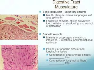
Structure of the wall in the GI tract • Mucosa • Epithelium (1) • Lamina propria (2) • Muscularis mucosae (3) • Submucosa (4)(submucosal plex.) • Muscularis externa (myenteric plex.) • Inner circular (5) • Outer longitudinal (6) • Adventitia / Serosa (7)
Esophagus • Non-keratinized stratified squamous epith. • Cardia-glands in the lamina propria • Muscularis mucosae is longitudinal • Glandulae esophageae in submucosa • Muscularis externa • Adventitia in the thoracic cavity, serora in the abdominal cavity
Stomach • Simple columnar epith. Gastric pits • Lamina propria: long, tubular glands • Mucous secreting neck cells • Chief cells on the base – pepsinogen • Parietal cells – gastic acid • Muscularis mucosae • Inner circular&outer longitudinal layers • Submucosa • Muscularis externa • 3 layers: inner obilque, middle circular, outer longitudinal • Serosa
Histology of the small intestine Structures helping absorbtion (with surface enlargment): • Circular folds(plicae circulares): More prominent in the duodenum and in the proximal part of the jejunum. They disappear at the terminal part of the ileum. • Villus (villi intestinales): projecting parts of the mucosa. They are more numerous in the duodenum and in the jejunum. Size: 0,5-1,5 mm • Microvilli: Projections on the apical surface of the epithelial cells. 3 1 2
Histology of the small intestine villi Lieberkühn cripts (intestinal galands) epithelium lamina propria Mucosa muscularis mucosae Submucosa circular longitudinali Muscularis externa Serosa
Ileum Paneth cells
58. Lung (HE) bronchus cartilage gland smooth muscle bronchiolus pseudostratified columnar epithelium alveolus
62. Stomach (HE) tunica mucosa tunica submucosa muscularis externa parietal cell chief cell
99. Ileum (HE) intestinal villus Lieberkühn's crypt tunica mucosa tunica submucosa Peyer's patch muscularis externa tunica serosa