Download

1 / 45
460 likes | 685 Views
Laboratory Test Results. Brenda C. Barnes and Shawn Froelich. Objectives. Correlate urinalysis reagent strip results with microscopic test results. Correlate microscopic evaluation results of a vaginal swab with clinical symptoms.

E N D
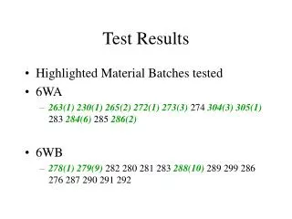
Laboratory Test Results Brenda C. Barnes and Shawn Froelich
Objectives • Correlate urinalysis reagent strip results with microscopic test results. • Correlate microscopic evaluation results of a vaginal swab with clinical symptoms. • Discuss proper collection procedures for anaerobic cultures. • Interpret microbiology susceptibility reports. • Explain the procedure for microscopic examination of skin scrapings. • Describe rapid testing available that is applicable to the clinical setting.
Chemical Exam – Glucose • Presence of glucose indicates the amount blood glucose has exceeded tubular reabsorptive capacity • Clinical correlation • Diabetes mellitus • Pancreatitis • Hyperthroidism • Gestational diabetes
Glucose • Clinitest • Nonspecific test for reducing substances • If performed, done so on pediatric specimens (< 2 years old) • Screening for galactosemia – may be part of state mandated newborn screening program • Microscopic Correlation • No elements seen • Yeast may be present
Chemical Exam – Bilirubin • Presence indicates liver disease or biliary obstruction • False-positives due to urine pigments • Ictotest – confirmatory test • Less subject to interference • False-negatives • Age of specimen – bilirubin is unstable • No microscopic correlation
Chemical Exam – Ketones • Presence indicates increased fat metabolism • Clinical significance • Diabetic acidosis • Insulin dosage monitoring • Starvation • Malabsorption • Microscopic correlation - none
Chemical Exam – Sp. Gravity • Strip reading is adequate for routine screening • Clinical significance • Monitoring patient hydration and dehydration • Loss of renal tubular concentrating ability • Diabetes insipidus • Determination of unsatisfactory specimens • Microscopic correlation – none
Chemical Exam – Blood • Presence of red blood cells, hemoglobin, or myoglobin • Hematuria - bleeding • Hemoglobinuria • Lysis of rbc in specimen • Intravascular hemolysis • Myoglobinuria – muscle destruction • Microscopic correlation – rbc seen in hematuria
Chemical Exam – pH • Of little diagnostic value – primarily used for determining systemic acid-base disorders • Microscopic correlation – none
Chemical Exam – Protein • Presence indicates abnormality in glomerular filtration barrier – renal disease • Correlates with: • Nitrite • Leukocytes • Microscopic
Chemical Exam - Urobilinogen • Increased in any condition that causes an increase in production or retention of bilirubin • Clinical significance • Early detection of liver disease • Lever disorders, hepatitis, cirrhosis, carcinoma • Hemolytic disorders • Microscopic correlation – none
Chemical Exam – Nitrite • Rapid screening for UTI • Sample needs to be fresh to avoid false-positive reactions • Correlates with: • Protein • Leukocytes • Microscopic
Chemical Exam – Leukocytes • Screening test for presence of wbc in urine • Quantification should be done by microscopic examination • Correlates with: • Protein • Nitrite • Microscopic
Microscopic Examination • Detect and identify insoluble materials present in urine • Time-consuming = cost • Lacks standardization Protocols used by many labs to improve standardization and cost-effectiveness
Vaginitis • Occurs when the mucosal lining of the vagina becomes inflamed and irritated • Typical signs: • Vaginal discharge • Vulvar itching irritation • Odor • Commonly associated diseases: • Bacterial vaginosis • Trichomoniasis • Candidiasis
Laboratory Diagnosis • Vaginal pH • KOH Amine “Whiff” test • Vaginal microscopy (wet mount) • Kit testing • BD Affirm • QuickVue Advance pH an Amines test • QuickVue Advance G. vaginalis test • OsomTrichomonas Rapid Test
Sample Collection • Swab vaginal vault and walls with one or two swabs • Include any areas where fluid has pooled • Place swab(s) in test tube containing 0.5 mL saline • Sample should remain at room temperature and tested within two hours of collection http://www.acponline.org/running_practice/mle/wm_exams.htm
Vaginal pH • Typical vaginal pH = 4.0-4.5 • > 4.5 • BV • Trichomoniasis • Tested at time of collection
Wet Mount • Vigorously mix swab(s) in and out of saline – collect all material adhering to side of tube • Remove swab from saline and depress onto clean, dry microscope slide – express small amount of fluid • Coverslip and examine under microscope http://www.acponline.org/running_practice/mle/wm_exams.htm
KOH “Whiff” Test • Prepare wet mount slide as directed, adding one drop of 10% KOH to slide prior to coverslipping • Positive test demonstrates typical “fishy” odor
Anaerobic Specimens • Most anaerobic infections are caused by endogenous microbiota • Improper collection may result in the growth of many anaerobes, resulting in difficulty to determine the cause of infection • Labs follow criteria for rejection of inappropriately collected and/or transported specimens
Acceptable Specimens for Anaerobic Culture • Aspirated material • CSF, blood, bone marrow, synovial fluid • Aspiration of closed abscess, ascites fluid, peritoneal fluid • Deep tissue or bone biopsy • Aspirated pus from decubitus ulcers • Suprapubic bladder aspiration • Pleural fluid obtained by thoracentesis, open lung biopsy, “sulfur granules” from draining fistula
Unacceptable Specimens for Anaerobic Culture • Swabs • Throat, nasopharyngeal, gingival, rectal, vaginal, cervical, urethral, surface wounds and abscesses • Expectorated or suctioned sputum, bronchial washings • Contents of large bowel, feces, colostomy effluents, gastric and small bowel contents • Voided or catheterized urine
Transport and Processing of Anaerobic Specimens • Transport and processing should be quick to maintain temperature, avoid exposure to oxygen and avoid dessication • Oxygen-free transport tubes/vials such as PRAS media (prereduced, anaerobically sterilized) • Anaerobic bags or pouches if delays in transport • Blood cultures require aseptic collection with bactericidal agent such as tincture of iodine or chlorhexidinegluconate with 70% alcohol to minimize contamination with normal skin biota
Reasons for Performing Antimicrobial Susceptibility Testing (AST) • Bacterial isolate is presumed to be the cause of infection and has unpredictable antimicrobial susceptibility • Patient allergies to 1st line agent • Example: allergy to penicillin • Provide alternative drug choices for treatment • Clinical response not as expected • Consider alternative explanations for lack of response; ex: S. pyogenes pharyngitis and penicillin- no resistance yet detected, but drug may not be reaching crypts of tonsils • Detection of emerging resistance (which may develop over course of therapy)
Reasons for Not Testing & Not Reporting Antimicrobial Susceptibility • Not performed when pattern is predictable for all strains (known susceptibility or resistance) • Streptococcus pyogenes(Group A strep) = S to penicillin • Antimicrobial agent does not accumulate at site of infection • Nitrofurantoin excreted in urine; does not reach therapeutic concentrations at sites other than urinary tract • In vitro testing does not reliably predict resistance • Cephalosporins and low dose aminoglycosides in Enterococcus; results may be misleading • Not performed on organisms considered normal flora for their common anatomical site • Exception: immunocompromised host
CLSI and FDA Standards in AST • Clinical and Laboratory Standards Institute (CLSI) publishes standards for AST testing and interpretation • CLSI suggests lists of FDA-approved antimicrobial agents for consideration and routine testing/reporting • FDA package inserts should be consulted for drug dosing and indications • FDA and CLSI are involved in developing MIC and zone diameter breakpoints • “Breakpoint” (cutoff) is the concentration of antibiotic agent that separates susceptible from intermediate or resistant results
Dilution Method- MIC TestOrganism suspension uses broth dilutions of antibiotic
MIC Testing Three results possible (as recognized by CLSI): S=Suceptible-organism should respond to usual doses administered by appropriate route I=Intermediate-isolate may be inhibited by concentrations achieved when maximum parenteral doses are given; may be selected, but consideration should be given to other choices that may provide optimal therapy R=Resistant-organism is not inhibited by achievable concentrations; drug should not be selected for therapy (except for certain body fluids where high concentrations of agent may accumulate
Disk Diffusion Test (Kirby Bauer)Organism tested with antibiotic disks, zones measured
Interpretative Standards for Zone DiametersThree results possible: S, I, R
Lab Reporting of AST Results • Objectives • Most clinically appropriate agent • Only agents appropriate for infection should be reported • Most cost effective • Least toxic • Consider host status and drug properties • Prevent resistance to more costly broad spectrum agents • Use of narrow spectrum agent prior to extended spectrum • Generally, Group A agents (narrow spectrum) reported before secondary agents • Group B agents reported if: • Isolate resistant to primary agents • Patient can’t tolerate primary agent • Secondary agent a better clinical choice or polymicrobial infection (more effective) *Exception: Meningitis- 3rd generation agents better at crossing blood-brain barrier
Example of Selective Reporting Protocol for E. coli and Enterobacter Primary agents are shown in plain print, and secondary agents are shown in bold print
Current Lab Challenges in AST • Oxacillin (methicillin) resistant Staphylococcus aureus(MRSA) • Vancomycin resistant Staphylococcus aureus(VRSA) or diminished susceptibility • Inducible clindamycin resistance in staphylococci • Vancomycin resistant enterococci (VRE) • Extended spectrum β-lactamases (ESBLs) • Klebsiellapneumoniaecarbapenemase (KPC) • Multiple drug class resistance in Streptococcus pneumoniae
Summary • In reporting susceptibility test results to clinicians, most labs will report only S or R, unless more data is needed • There are many complex factors in AST, and increasing resistance patterns are emerging • Choice of antibiotic should start with the most clinically effective, cost effective, and least toxic, to prevent further organism resistance • In vitro AST results may not correlate directly with in vivo action due to numerous pharmacokinetic and pharmacodynamic factors • Always use proper infection control (handwashing) to prevent spread of community-acquired and nosocomial pathogens (especially emerging resistant strains)
References Clinical Laboratory Standards and Institute. Available from: http://www.clsi.org/ Marsik, Frederic J., (2011). Antimicrobial Susceptibility Testing. Mahon, Connie R., Lehman, Donald C., Manuselis, George. In Textbook of Diagnostic Microbiology (4thed). (pp. 276-307). Maryland Heights: Saunders Elsevier. Winn, Jr. Washington, Allen, Stephen, Janda, William, Koneman, Elmer, Procop, Gary, Schreckenberger, Paul & Woods, Gail. (2006). Antimicrobial Susceptibility Testing. InKoneman’s Color Atlas and Textbook of Diagnostic Microbiology (6thed). (pp. 945-1021). Baltimore: Lippincott Williams & Wilkins.