Download
1 / 31
350 likes | 993 Views
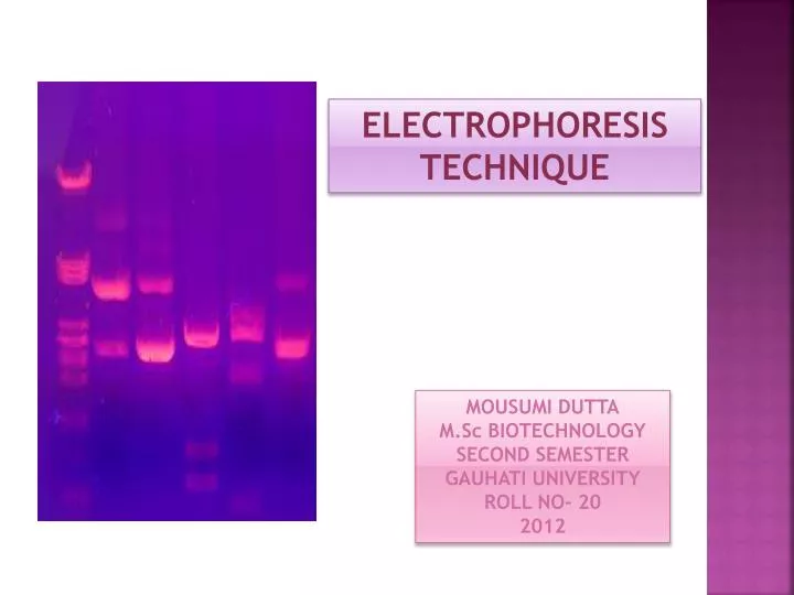
ELECTROPHORESIS TECHNIQUE. MOUSUMI DUTTA M.Sc BIOTECHNOLOGY SECOND SEMESTER GAUHATI UNIVERSITY ROLL NO- 20 2012. electrophoresis. The history of electrophoresis begins with the work of Arne Tiselius in the 1930s.

E N D
ELECTROPHORESIS TECHNIQUE MOUSUMI DUTTA M.Sc BIOTECHNOLOGY SECOND SEMESTER GAUHATI UNIVERSITY ROLL NO- 20 2012
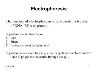
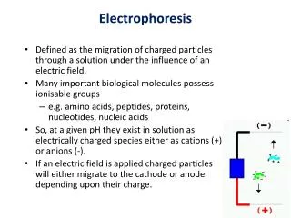
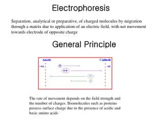
electrophoresis The history of electrophoresis begins with the work of Arne Tiselius in the 1930s. Electrophoresis is a separations technique that is based on the mobility of ions in an electric field. In order words it is a technique for separating molecules in a mixture under the influence of an applied electric field. Dissolved molecules in an electric field move, or migrate, at a speed determined by their charge : mass ratio.
TYPES- • Gel Electrophoresis • Pulsed field gel electrophoresis • SDS- Polyacrylamide Gel Electrophoresis • Two-Dimensional Gel Electrophoresis • Capillary Electrophoresis • Affinity Electrophoresis • Dielectrophoresis • Immunoelectrophoresis • Isotachophoresis
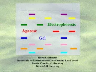
GEL ELECTROPHORESIS • Used in clinical chemistry to separate proteins by charge and ∕ or size. • In biochemistry and molecular biology to separate DNA and RNA fragments by length, size or to separate proteins by charge.
Separation based on- • Molecules are separated or sorted by applying an electric field to move the negatively charged molecules through an agarose matrix. • The molecules being sorted are dispensed into a well in the gel material placed in an electrophoresis chamber, connected to a power source. • When the electric current is applied, the larger molecules move more slowly through the gel while the smaller molecules move faster.
The different sized molecules form distinct bands on the gel. • Electrophoresis" refers to the electromotive force (EMF) that is used to move the molecules through the gel matrix. • Samples loaded into adjacent wells in the gel, run parallel in individual lanes. • Components from the original mixture separates as one or more distinct bands.
Type of Gel used- Agarose gel- • Used for separating larger nucleic acids ranging from 50 to million base pairs. • Distance between DNA band is determined based on the amount of agarose used. • But larger the concentration of agarose longer the time of run. Polyacrylamide- • Used for separating proteins ranging in size from 5 to 2,000 kDa. • Pore size is based on the concentrations of acrylamide and bis-acrylamide powder used in creating a gel. • Used to separate DNA fragments differing by a single base-pair in length.
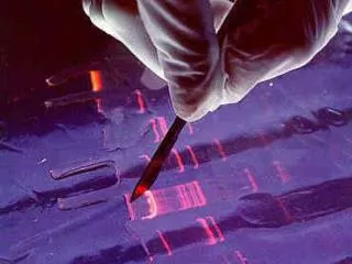
Buffersused- • The most common being, for nucleic acids Tris Acetate EDTA(TAE) and Tris Borate EDTA(TBE). • TAE has the lowest buffering capacity but provides the best resolution for larger DNA. Visualization- • The molecules in the gel can be stained to make them visible. • DNA may be visualized using ethidium bromidewhich, when intercalated into DNA, fluoresce under ultraviolet light. • Photographs can be taken of gels, often using Gel Doc.
Pulsed field gel electrophoresis • Used for separation of large DNA molecules by applying an electric field that periodically changes direction to a gel matrix. • Procedure takes longer than normal gel electrophoresis due to the size of the fragments being resolved and that DNA does not move in a straight line. • Various lengths of DNA reacts to the change at differing rates. • Used for genotyping or genetic fingerprinting
SDS Polyacrylamide gel electrophoresis • Polyacrylamide gel electrophoresis(PAGE) provides versatile, gentle, high resolution for fractionation and physical separation of molecules on the basis of size, charge. • Used for separating proteins of smaller size due to uniform pore size of the gel. • SDS(sodium dodecylsulfate) denatures proteins, causing multimeric proteins to dissociate into their subunits, and thus separated on charge : mass ratio. • Even chains that differ in molecular weight by less than 10 percent can be separated by this technique.
TWO DIMENTIONAL GEL ELECTROPHORESIS • Commonly used to analyze proteins. • Separated by two properties in two dimensions on 2D gel. • Begins with 1D electrophoresis but then separates the molecules by a second property. • Firstly separated by their charges(isoelectric point) and then by their masses.
Gel strip contains a continuous pH Gradient formed by ampholytes, a mixture of polyanionic and polycationic molecules . • A charged protein will migrate through the gradient until it reaches its isoelectric point (pI), the pH at which the net charge of the protein is zero. • Molecules are thenseparated in a second dimension on the basis of their molecular weights. • IEF gel separates molecules according to charges and SDS gel separates molecules on the basis of masses. • Useful in comparing the proteomes in undifferentiated and differentiated cells.
Capillary electrophoresis • Also known as Capillary zone electrophoresis used to separate ionic species by their charge and frictional forces and hydrodynamic radius. • Two different principal types of separation matrix are used- • high viscosity polymer- polyacrylamide. • low viscosity polymer- agarose. • The velocity of migration of an analyte depends upon the rate of electro osmotic flow of the buffer solution. • Analyte migrate towards the electrode of opposite charge.
Fused silica capillaries forms the capillary surface. • CE uses a very high voltage (1–30 kV) for the separation of analytes in the capillary, which may be either coated internally, or uncoated. • Phosphate buffers are generally used. • By adjusting the pH of the buffer, the electro osmotic flow can either enhance or oppose electrophoretic migration.
The ionization of the capillaries can be enhanced by running a basic solution such as NaOH or KOH. • Capillary isoelectric focusing (CIEF) utilizes ampholytes that span the pH range of interest. • Applications include DNA diagnosis of Down’s syndrome (25), adenylosuccinate lyase deficiency (26), and P53 oncogene analysis.
AFFINITY ELECTROPHORESIS • It includes mobility and charge shift electrophoresis and affinity capillary electrophoresis. • It includes complex formation between macromolecules and the membrane proteins. • The binding of a molecule normally changes the electrophoretic properties of a molecule. • Characterization of molecules with specific features like glycan content or ligand binding.
DIELECTROPHORESIS • It is a phenomenon in which a force is exerted on a dielectric particle when it is subjected to a non-uniform electric field. • It does not require the particles to be charged. • Depends on the medium and particle’s electric properties, shape, size etc. • Example- Separation of cells or orientation and manipulation of nanoparticles.
IMMUNOELECTROPHORESIS • Involves separation and characterization of proteins based on electrophoresis and reaction with antibodies. • It requires immunoglobulins reacting with proteins to be separated. • Agarose gel is used. • Immunoprecipitates can be seen on staining with protein stains such as- Coomassie Brilliant Blue.
ISOTACHOPHORESIS • Used to separate charged particles. • Uses a discontinuous electric field to create a sharp boundaries between the sample constituents. • Sample is introduced between a fast leading electrolyte and a slow terminating electrolyte. • Constituents are separated from each other by sharp electric field differences. • Cerebrospinal fluid analysis(CSF) in the diagnosis of multiple sclerosis uses IEF.
References- • Keith R. Mitchelson Jing Cheng; Capillary electrophoresis of nucleic acids. • Molecular cell biology; Lodish. • Molecular biology of the cell; Alberts. • Garrett and Grisham; Biochemistry. • wikipedia.