Download
1 / 4
40 likes | 54 Views
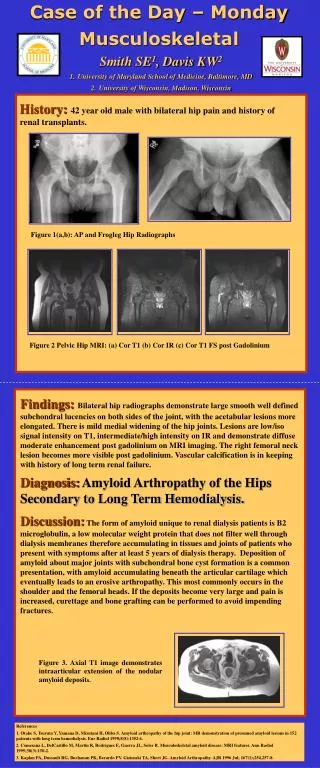
Sunday Case of the Day. Physics. Authors: Lingyun Chen Ph.D., Lifeng Yu, Ph.D., Shuai Leng, Ph.D., Cynthia McCollough, Ph.D. Department of Radiology, Mayo Clinic, Rochester MN. History: A 55 years old male patient underwent a contrast-enhanced abdomen and pelvis MDCT scan.

E N D
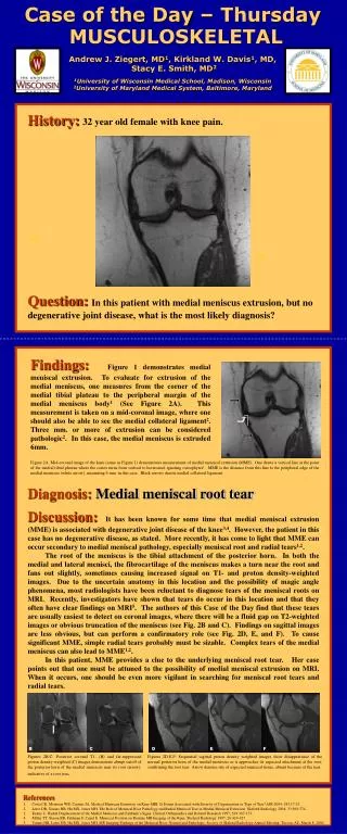
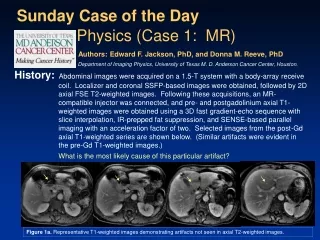
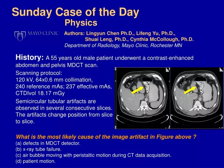
Sunday Case of the Day Physics Authors:Lingyun Chen Ph.D., Lifeng Yu, Ph.D., Shuai Leng, Ph.D., Cynthia McCollough, Ph.D. Department of Radiology, Mayo Clinic, Rochester MN History: A 55 years old male patient underwent a contrast-enhanced abdomen and pelvis MDCT scan. Scanning protocol: 120 kV, 64x0.6 mm collimation, 240 reference mAs; 237 effective mAs, CTDIvol 18.17 mGy Semicircular tubular artifacts are observed in several consecutive slices. The artifacts change position from slice to slice. What is the most likely cause of the image artifact in Figure above ? (a) defects in MDCT detector. (b) x-ray tube failure. (c) air bubble moving with peristaltic motion during CT data acquisition. (d) patient motion.
Diagnosis:The correct answer is (c). The cause of this artifact is that gas bubbles moving in the stomach during the image acquisition process.
Discussions • The gas bubble motion artifact is an arc structure with an air attenuation level that results from reconstruction of the CT data acquired during the motion of a gas bubble. The severity of this artifact depends on the size and velocity of gas bubble, scanning speed and relative position of the x-ray tube. • Liu F. et al performed a CT scan of a water filled body phantom with simulated gas bubble motion inside. A tube was affixed to the bottom of the phantom and a CT injector was filled with 60 ml room air to inject air into the tubing at a controlled rate. The phantom was scanned with clinical routine protocol on a 16-MDCT scanner and reconstructed with same parameters as for the patient images. Figure: Motion artifacts caused by moving gas bubble are demonstrated in this phantom experiment. Air injection rate is 0.1 ml/s (left) and 0.5 ml/s (right). (see ref. 2) • The artifact could obscure anatomic structures and generate non-diagnostic images. When the artifact is subtle, it could simulate an abnormality which may lead to wrong diagnosis. • It is difficult to completely eliminate the presence of a moving gas bubble in the patient during a CT scan. Any method to reduce the chance of generating moving gas bubbles is helpful. In terms of scanning technique, improving the temporal resolution or the scanning speed is also helpful to reduce the motion artifact generated by moving gas bubble if it is present.
References • J. Hsieh, Computed tomography: principles, design, artifacts, and recent advances. Second edition, Bellingham, WA: SPIE Press, 2009. • F. Liu et. al, Gas Bubble Motion Artifact in MDCT. AJR 2008; 190:294–299. • CH McCollough et al, Motion artifacts in subsecond conventional CT and electron-beam CT: pictorial visualization of temporal resolution. RadioGraphics 2000; 20:1675-1681