Download
1 / 44
450 likes | 817 Views
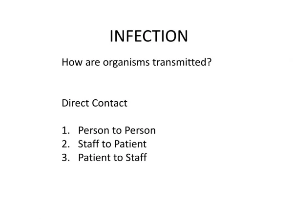
Infection. 2006 Dr. Nasser Rizk. Infection. Invasion and multiplication of microorganisms inside body producing S&S and immune response. Severity of infection depends on : 1- Pathogenicity 2- number 3- host defenses Transmission of infection needs:

E N D
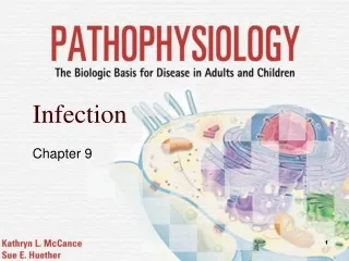
Infection 2006 Dr. Nasser Rizk
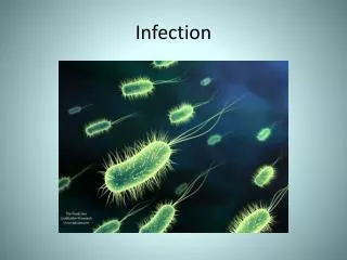
Infection • Invasion and multiplication of microorganisms inside body producing S&S and immune response. • Severity of infection depends on : 1- Pathogenicity 2- number 3- host defenses • Transmission of infection needs: 1- Causative agent 2- reservoir 3-port of transmission 4-susceptible host
Risk Factors • 1-Weak defense mechanisms • 2- Enviromemental factors • 3- Developmental factors • ----------------------- • Weak Defense mechanisms: • Immunodeficiency or Immunocompromise • Impaired WBCs and low level of T and B cells (immunodeficiency) • May be congenital (before or at birth) or acquired (after birth) • body’s ability to recognize and fight pathogens is impaired
Risk Factors (continued) • Conditions which suppress immune Response: 1-Diabetes mellitus 2 –Renal failure 3- Liver cirrhosis 4- Steroids 5-Immunosuppresive drugs (transplantation) 6- Chemotherapy. Acquired immune suppression in: stress malnutrition infections pregnancy
2- Enviromemental factors • Conditions weaken a person immune defense are: 1- Poor hygiene 2- Malnutrition 3- Inadequate barriers 4- Stressors 5- Chronic diseases 6- Inadequate treatment 7-Inadeqaute immunization Dust facilities transport of pathogens, e.g., Aspergillus, (lung)
3- Developmental factors • Children and old age at high risk. • Children : immune system is not fully developed (6 months) • most common: respiratory infections • Exposure to communicable diseases is high in care- centers and schools. • Old age: decline in immune system • DM and atherosclerosis: weak delivery of nutrients by impaired blood flow to organs
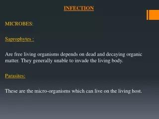
Components of the chain of infection • Agents: • Bacteria, Viruses, • Fungi, Parasites, Mycoplasma, • Ricketessia, Chlamydia • Reservoir: • Microbes can survive • E.g., humans, animals , etc
Components (continued) • Portal of exit (entry): • infectious agent leave (enter) the organism • Respiratory, genitourinary, GIT, skin, Mucus membranes, and placenta. • Secretions as : blood, sputum, emesis, stools, urine, wound drainage and genital secretions act as portal. Mode of transmission Means from portal of exit to host 4 modes: chart 3 Vector- born: flea, mosquito (tropical) Chart 3
Stages of infection • 1- Incubation: replication, transmission. • 2-Prodromal stage: vague complaint, transmission. • 3- Acute illness: microbes destroy host cells, affecting systems. • 4- Convalescent stage: body defense take over the microbe, healing.
Pathophysiologic changes • Characteristic changes: Prodromal stage 1- Fever 2- Muscle aches 3- Headache 4- Lethargy Acute stages: Moe specific symptoms
Inflammation • A major reactive defense mechanism against infective agents. • It may result from: 1- tissue injury 2- infection 3- allergy • Stages: • 1- Vascular 2- Cellular Vascular: arterioles constrict then dilate--------edema Cellular: inflamm. Cells release histamine---edema Which dilute microbes
Signs and Symptoms of Inflammation • Redness • Heat • Pain • Edema • Loss of function
These were known Heat Redness Swelling Pain Loss of function by the Romans Calor Rubor Tumor Dolor Functio laesa Five classic local signs of acute inflammation Added Later
Five classic local signs of acute inflammation • The major components responsible for these local signs are • Heat - vasodilatation • Redness - vasodilatation • Swelling - vascular permeability • Pain - mediator release/pmn’s • Loss of function - mediator release/pmn’s
Alerts !! • 1- Localized infections: • produce rapid inflammatory response • Obvious symptoms and signs • 2- Disseminated infections: • Slow inflammatory response • Long time for treatment
Inflammation • Acute - minutes to days • Characterized by fluid and protein • PMN’s • Chronic - weeks to years • Lymphocytes and macrophages • ACUTE Inf - PMN’s (Polymorphonuclear Cells) • CHRONIC Inf - Mononuclear Cells EXUDATE
Fever • After introduction of infectious agent • Elevated temperature helps to fight infection • Many organisms can’t survive • Diaphoresis is the method of cooling down • Improve immune system rspone.
Leukocytosis • Body’s response to introduction of pathogens; WBCs increases • Neutrophils count increases, with increase immature cells “bands”, but not function • Neutrophils, monocytes, and macrophages begin phagocytosis of dead tissues and bacteria • They identify the foreign antigen and kill the microbe • Elevated count of WBCs is common; N in acute stages and Monocytes during resolution or chronic stages
Chronic inflammation • Infection longer than 2 weeks • May occur after acute Inflammation • Permanganate scarring and loss of function may occur • E.g., Mycobacterium tuberculosis
Diagnosis • Medical history • Examination • Investigations-tests • WBCs and CBC (first test) • ESR: increased, inflammatory response • Gram stain, silver stains • Culture- sensitivity tests • MRI to locate infection sites • Chest X-ray (lung) • Gallium scan for abscess detection
Treatment • Vary widely • Use vaccines----1ry immune response • Drugs when appropriate • Supportive therapy • Antibiotics, antiviral, antifungal----- • Overuse of medication----resistance • Proper prevention of epidemic and pandemic transmission.
Acute inflammation • The immediate and early response to injury • The point? Get the pmn’s to the site as fast as possible • Vasodilatation • Endothelial permeability • Extravasation of pmn’s
Acute inflammation major components • Vasodilatation • Endothelial permeability • Extravasation of pmn’s
Vascular changes you need to know this • Vasodilation (forget the few seconds of vasoconstriction) • Exudation of protein rich fluid • Blood stasis • Margination • Emigration/Transmigration
Vascular changes you need to know this • Vasodilation (forget the few seconds of vasoconstriction) • Exudation of protein rich fluid • Blood stasis • Margination • Emigration/Transmigration
Vascular permeability • Vasodilation, increased blood flow • Increased intravascular hydrostatic pressure • Transudate - ultrafiltrate blood plasma (contains little protein) • Again, this is very transient and just gets the process started. Think Acute Inflammation, think EXUDATE • Increased vascular permeability
Vascular permeability • Exudate - (protein-rich with pmn’s) • Exudate is the characteristic fluid of acute inflammation • Intravascular osmotic pressure decreases • Osmotic pressure of interstitial fluid increases • Outflow of water and ions - edema
How do endothelial cellsbecome leaky? • Endothelial cell contraction • Junctional retraction • Direct endothelial injury (immediate sustained response) • Leukocyte-dependent endothelial injury • Increased transcytosis of fluid
Direct endothelial injury (immediate sustained response) • Endothelial cell necrosis and detachment • Result of severe injury or burn • Occurs immediately and lasts until vessel repaired
Leukocyte-dependent endothelial injury • Occurs at sites of leukocyte accumulation • Due to leukocyte activation which releases proteolytic enzymes and toxic oxygen
Leukocyte Cellular Events • Margination and Rolling • Adhesion and Transmigration • Migration into interstitial tissue
Margination • Normal flow - RBC’s and WBC’s flow in the center of the vessel • A cell poor plasma is flowing adjacent to endothelium • As blood flow slows, WBC’s collect along the endothelium • This is “Margination”
Endothelial Activation • The underlying stimulus causes release of mediators which activate the endothelium causing selectins and other mediators to be moved quickly to the surface
Selectins • Selectins bind selected sugars • Selected + Lectins (sugars) = Selectins • Some selectins are present on endothelial cells (E-Selectin) • Some selectins are present on leukocytes (L-Selectin) • Some selectins are present on platelets (P-Selectin)
Chemotaxis • Movement toward the site of injury along a chemical gradient • Chemotactic Factors include • Complement components • Arachadonic Acid (AA) metabolites • Soluble bacterial products • Chemokines
Leukocyte Activation • Chemokines also “activate” PMN’s • AA metabolite production • Degranulation and Secretion of lysosomal enzymes • Oxidative burst • Modulation of adhesion molecules
Phagocytosis & Degranulation • Phagocytosis (to eat and destroy) • Attach • Engulf • Kill • Degranulation and the oxidative burst destroy the engulfed particle
Leukocyte-induced tissue injury • Lysosomal enzymes are released into the extracellular space during phagocytosis causing cell injury and matrix degradation • Activated leukocytes release reactive oxygen species and products of arachidonic acid metabolism which can injure tissue and endothelial cells • These events underlie many human diseases (e.g. Rheumatoid arthritis)
Pneumonia Another picture of the same thing… At a higher power!