Download
1 / 16
240 likes | 795 Views
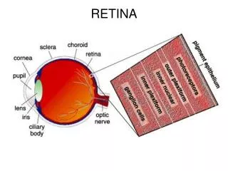
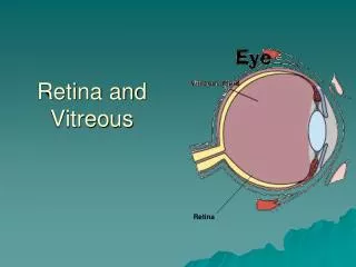
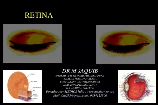
Photoreceptors and Retina. I. Structure of retina - The retina is composed of three main layers 1) Outer nuclear layer: photoreceptors, no direct blood supply 2) Inner nuclear layer: bipolar, horizontal, amacrine , Mueller glial cell bodies .

E N D
I. Structure of retina • - The retina is composed of three main layers • 1) Outer nuclear layer: photoreceptors, no direct blood supply • 2) Inner nuclear layer: bipolar, horizontal, amacrine, Mueller glial cell bodies.
3) Ganglion cell layer: retinal ganglion cells • - The outer and inner plexiform layers are sites of synapses between layers
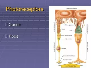
II. Photoreceptors • - There are two types of photoreceptors • 1) rods: most sensitive in dim light, only one type • 2) cones: less sensitive to light, three types mediate color vision.
- Distribution of photoreceptors is not uniform: • fovea has high density of cones and few rods, periphery has more rods. • - The outer segment of rods and cones have stacks of disks with high density of pigment and provide a high probability of absorption of an incident photon.
- Pigment epithelial cells serve to • (1) absorb light not absorbed by the rods/cones using melanin, • (2) phagocytoseshedded fragments of rods/cones, • (3) regenerate pigment
III. Visual pigments • - Pigment is composed of a chromophore covalently bound to an opsin 7TM protein • - Humans have 4 pigments (1 rod, 3 cones) each maximally sensitive at different wavelengths.
- Upon photon absorption, the chromophore undergoes several configuration changes; • the configuration that triggers vision takes several ms to reach; • the ultimate result is the release of chromophore, and binding of fresh chromophore with opsin.
IV. Phototransduction • - Rods/cones hyperpolarize in response to light in a graded fashion with respect to intensity, with cones requiring more light and responding more rapidly • - Phototransductiona prototypical signal transduction mechanism.
- In dark, cGMP keeps a nonselective cation channel open, and cell is depolarized • - Rhodopsin is coupled to G-protein that activates phosphodiesterase and lowers [cGMP] • - Ca influx plays a key negative feedback function and mediates light adaptation.
V. Synaptic connections in the retina. • - Throughput pathway: photoreceptor bipolar retinal ganglion cell optic nerve • - Lateral associations: between photoreceptors, and via horizontal and amacrine cells • - There are distinct synapse morphologies in the retina.
- Synaptic triad: photoreceptor with horiztonal-bipolar-horizontal cells, and contains a synaptic ribbon • - Dyad: proximal bipolar end synapses with two cells • - Primary nxt’s in the retina are Glu (excitatory) and GABA (inhibitory).
VI. Information processing in the retina • - The receptive field of a single photoreceptor is bigger than itself due to connections with neighboring photoreceptors; • for the same reason, the receptive field of horizontal cells is bigger than the multiple photoreceptors it contacts.
- Since bipolar cells receive synapses from both photoreceptors and horizontal cells, their receptive field is divided into a center (dominated by photoreceptor) and surrounding ring (dominated by horizontal cells) • - In light, on-bipolars depolarize in response to light in the center of its receptive field and hyperpolarize in response to light on the surrounding ring; off-bipolars are vice versa.
- The above property of bipolars, propagated to on- and off-center ganglion cells, probably helps mediate contrast and edge detection • - Ganglion cells can also be classified as X (slow adapting) and Y (fast adapting) types • - Amacrine cell firing pattern is best fit to respond to moving objects.
VII. Photoreceptors for photoentrainment (will not be tested) • - A small percentage of ganglion cells are melanopsin positive, these project to regions of the brain responsible for photoentrainment and pupil reflex (e.g. the suprachiasmatic nucleus) • - It has not been proven definitively that melanopsin is actually a photoreceptive pigment—yet.