Download
1 / 52
520 likes | 574 Views
Fatal Familial Insomnia: Pathogenesis caused by a mutation affecting the metabolism of the normal prion protein. By Sabrina T. Gillig BIO-475 Seminar Dr. Peter Lin. FATAL FAMILIAL INSOMNIA the nightmare of those who never sleep.

E N D
Fatal Familial Insomnia: Pathogenesis caused by a mutation affecting the metabolism of the normal prion protein.By Sabrina T. GilligBIO-475 SeminarDr. Peter Lin
FATAL FAMILIAL INSOMNIAthe nightmare of those who never sleep • Dateline NBC. 2005. Fatal Insomnia: Genetic mutation inflicts rare disease through generations. http://www.msnbc.msn.com/id/6822468/ . Accessed 11 APR 2006. CLICK ON THE LINK TO WATCH THE VIDEO !
What are Prions? • Prions are the smallest infectious particles known to date. They are made only of a protein. • Prions are abnormally folded proteins. • Prions are the cause of transmissible spongiform encephalopathies. • Prion diseases are fatal and untreatable. Cann, 1997.
More on prions • The normal prion protein PrPc is found primarily on the surface of neurons, and is likely to be a synaptic protein with functional role in the synaptic transmission. • Prion diseases exhibit an extended latency period, spending this time performing neuroinvasion.
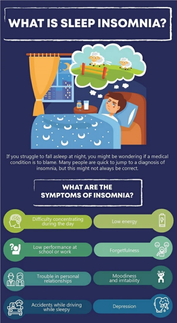
What is Fatal Familial Insomnia? • FFI is an autosomal dominant inherited disease; cause by a mutation of the normal prion protein. • A mutation in codon 178 replaces asparagine (N) for aspartic acid (D)→ D178N • First symptoms to arise trouble sleeping, difficulty concentrating , and personality changes • These symptoms usually appear during midlife, after appearance of the first symptoms death follows usually with 18 months.
The Thalamus (Wikipedia, 2006) • The thalamus is the point where most signals from the CNS pass to the cerebrum. • Severe loss of neurons in the thalamic nuclei, and accumulation of amyloid plaques. • Sponge-like appearance under the microscope-Transmissible Spongiform Encephalopathies (TSEs)
Treatment • There is no treatment for FFI. • Two drugs (quinacrine and chlorpromazine) were being tested, but the individuals in the clinical trials worsened.
Based on… Petersen, R.B., P. Parchi, S.L. Richardson, C.B. Urig, and P. Gambetti. 1996. Effect of the D178N mutation and the codon 129 polymorphism on the metabolism of the prion protein. The Journal of Biological Chemistry 271: 12661-12668.
Metabolism of the mutated prion protein • Codon 129 Polymorphism • PRNP encodes for methionine or valine. • CJD and FFI similarities • The normal haplotype is designated 178D, the haplotype in FFI is designated D178N.
The metabolic and mutational events that lead to the syndrome will be examined further along this presentation…
Expression, Localization and metabolism of the PrP in humans • PrPc is a glycoprotein attached to the cell membrane . • During the process of translocation in the ER, PrPc continues its folding process. Further folding occurs in the Golgi apparatus (←See graph) ( Schuldiner et al., 2005)
N-Linked Glycosylation Is common to all eukaryotic cells. It is imperative for proper folding of the protein.
The secretory Pathway ( Schuldiner et al., 2005)FOCUS: N-LINKED GLYCOSYLATION
The Secretory Pathway and Protein Glycosylation • The secretory pathway is the method that cells use to transport different materials to the outside. • The synthesis of glycoproteins is a very complex process which takes place in three different cell compartments: in the cytoplasm, in the membrane of the endoplasmic reticulum (ER) and in the Golgi complex .
N-GLYCOSYLATION Step-by-step • Starts on the surface of the ER membrane by the addition of two N-acetylglucosamines and five mannoses. • Then, the whole complex is t flipped to the luminal side of the membrane, where four further mannoses and three glucoses are added. • As the dolichylphosphate is released, the whole oligosaccharide is transferred to the asparagine residues of polypeptide chains that are in the process of assembling into protein complexes. ( Schuldiner et al., 2005)
N-GLYCOSYLATION Step-by-step (Continued) • While the proper protein folding is taking place, the three glucoses and one mannose are trimmed away by numerous different enzymes . • Using vesicles, the glycoprotein is then transferred to the Golgi complex, where additional modifications take place. • Finally, the complex is completed with one fructose and three galactose- and sialic acid molecules by numerous different enzymes and exported to the outside.
Schematic Representation of The Prion Protein -The human PrP. Polymorphic sites 129 and 178 are shown. GPI (ground positioning indicator) anchors the prion protein to the cell membrane.
Three forms of PrPc • Called GLYCOFORMS and differ in the level of glycosylation (addition of sugar units). • 1) Unglycosylated • 2)monoglycosylated • 3)diglycosylated
Cell lines in this study were obtained from human neuroblastoma cells and inserted with the PrP sequence, either MET or VAL. Scientists wanted to evaluate the outcome of the polymorphism on the metabolism of the PrP. Cell lines and their different genotypes
The cells were treated with PI-PCL. →PI-PCL is used to unanchor the PrP from the cell membrane. Scientists were able to study the prion protein itself and quantify solely the amount of PrP present on the cell surface. PrPm amount leaded was 3 times higher than PrPc to further confirm its underrepresentation on the cell surface. What are the glycosylation differences among mutant and normal cells on the cell surface? M: mature I: Intermediate U: Unglycosylated
Results • 3 different glycoforms were observed • Only 1/3 the amount of PrPm was released from the cell surface of mutant cells when compared to the control.
Carbohydrates in the sample were removed by the enzyme N-Glycosidase. This helped scientists observe exclusively the amount of PrP present in normal and mutant cell surface according to their size. Glycosylation differences on the cell surface-Normal vs. Mutant-
Results • Theunglycosylated form is underrepresented on the cell surface.
To prove that the mutant prion protein is efficiently unanchored by the PI-PCL treated (+), and untreated (-) cells were labeled with biotin. As seen in this figure, PrPm is powerfully cut by PI-PCL treatment Efficiency of the PI-PCL on the mutant PrP
Scientists concluded that… • The three PrP forms differ in the level of glycosylation→ In mutant cells the unglycosylated form is virtually inexistent.
Scientists wanted to quantitate the amount of PrP present inside the cell and on the cell surface. Notes: -0-0.5-2 Hours: Different significant times along the Pulse chase study. M: Mutant I: Intermediate migrating form U: Unglycosylated Prion Protein representation inside the cell and on the cell surface
Graphical Representation of unglycosylated PrP intracellularly and on the cell surface. WM: wild methionineWV: wild valineMM: mutant methionine (FFI)MV: mutant valine (CJD)
Why? • Because the underrepresentation of PrPm on the cell surface could be the result of rapid turnover (degradation) or unsuccessful transport. • Also, it could be the result of selective degradation or increased glycosylation. SCIENTISTS WANTED TO KNOW WHAT IS HAPPENING TO THE PrP INSIDE THE CELL…
How did they do this? • PULSE CHASE By labeling the samples with radioactive material, they were able to quantitate the amount of protein present at each different time, and deduce its location.
What does this mean? • INTRACELLULARLY: At zero time PrP is still in the ER-Golgi complex • ON THE CELL SURFACE: The mutant PrP is inadequately transported during the secretory pathway;
Glycosylation was prevented using the antibiotic tunicamycin. Tunicamycin prevents glycosylation by blocking the synthesis of the high mannose core of newly synthesized proteins in the ER. -Tunicamycin treated (+) Untreated (-) How does the stability of PrPm affect its transport to the cell surface?
Results • The unglycosylated form of the D178N PrPm is degraded inside the cell, while the normal PrPc necessitates glycosylation to reach the cell surface. • When glycosylation is prevented, the PrPm hardly arrives to the cell surface, and untraceable after synthesis.
PrP Degradation • Experimental In order to find out whether PrPm is degraded when kept in the ER-Golgi compartment scientists used brefeldin A -which blocks transport of glycosylated proteins from the ER to the Golgi complex-. ( Schuldiner et al., 2005)
Scientists wanted to find out where the PrPm is being degraded.. The PrPc showed no major changes with time, all forms were easily detectable. PrPm amounts decreased indicating degradation or conversion to the glycosylated form. What are the effects of transport block in the degradation of the PrP?
Results • Normal cells + Brefeldin A All three glycoforms were observed • Mutant cells + Brefeldin A Mutant cells exhibited degradation or change to the glycosylated form →More unglycosylated PrPm reaches the cell surface when valine is present in codon 129.
Results (continued) • Degradation of the mutant prion protein does not occur in the Golgi compartment, but in the endosomal-lysosomal system, which contains highly acidic enzymes.
D178N mutant cells lack PrPres • Normal and mutant cells were tested for proteinase K-resistant PrP. • Mutant cells lack PrPres which provides resistance to powerful denaturing conditions.
Underrepresentation in the brain of the PrPm • Western Blot was used to determine whether the unglycosylated form of the mutant prion protein is decreased in FFI patients. • Portions of normal and diseased brain were examined.
Presence of unglycosylated PrP in the brain of FFI patients Scientists examined the PrPm present in a FFI patient, in a section of the brain which does not have PrPres. All N-linked sugars in the FFI patient sample were removed by the enzyme PNGase, to study the original glycoforms. Lane 1: Untreated normal Lane 2: Untreated mutant Lane 3: PNGase treated normal Lane 4: PNGase treated mutant Control lane 3/ FFI lane 4
Results • In the mutant gray matter the unglycosylated form is present at only about 1/3 when compared to the normal samples.
What appears to be the cause of disease in FFI? • FFI appears to result only from the degradation of the unglycosylated form before it reaches the cell surface.
Conclusion • Pathogenesis in FFI and other prion diseases is believed to be caused by a change in the shape of the normal protein. • It is imperative to continue research, since in other neurodegenerative diseases (e.g. Alzheimer's) a misfolded protein could also be the cause. • A detailed analysis of the different factors, mechanisms and disease expression may be critical in the event of an epidemic (Mad Cow disease in the mid 1990’s).
Even though FFI and other prion diseases are rare and sporadic, science should always try to stay a step ahead…for the sake of all humanity.
Resources • Aguzzi, A. 2004. Peripheral pathogenesis of prion diseases. Pp.145-189 in Prions and prion diseases. Horizon Bioscience, England. • Aguzzi, A., F. Montrasio, and P.S. Kaeser. 2001. Prions: health scare and biological challenge. Nat Rev Mol Cell Biol 2: 118-126. • Aguzzi, A., and C. Weissmann. 1997. Sleepless in Bologna: transmission of fatal familial insomnia. Trends in Microbiology 4: 129-131. • Benito-Leon, J. 2004. Combined quinacrine and chlorpromazine therapy in fatal familial insomnia. Clin. Neuropharmacol. 27: 201-203. • Blatter, T., S. Brandner, A.J. Raeber, M.A. Klein, T. Voigtlander, C. Weissmann, and A. Aguzzi. 1997. PrP-expressing tissue required for transfer of scrapie infectivity from spleen to brain. Nature 389: 69. • Bosque, P.J., C.L. Vnencak-Jones, M.D. Johnson, J.A. Whitlock, and M.J. McLean. 1992. A PrP gene codon 178 base substitution and a 24-bp interstitial deletion in familial Creutzfeldt-Jakob disease. Neurology 10: 1964-1870. • Butler, G.H., H. Kotani, L. Kong, M. Frick, S. Evancho, E.J. Stanbridge, and G.J. McGarrity. 1991. Identification and characterization of proteinase K-resistant proteins in members of the class Mollicutes. Infect Immun 59: 1037-1042. < http://www.pubmedcentral.nih.gov/articlerender.fcgi?artid=258364 >. Accessed 09 Mar 2006. • Cancerweb. 1997. Endosomes. < http://cancerweb.ncl.ac.uk/cgi-bin/omd?query=endosomes >. Accessed 09 Mar 2006. • Cann, A.J. 1997. Principles of molecular virology. Prion picture < http://www.triroc.com/sunnen/topics/prion.htm >. Accessed 07 Mar 2006. • Caughey, B., R.E. Race, D. Ernst, L.L. Perry, B. Chesebro, and R.E. Race. 1990. Normal and scrapie-associated forms of prion protein differ in their sensitivities to phospholipase and proteases in intact neuroblastoma cells. Journal of Virology 64: 1093. • Caughey, B., R.E. Race, M. Vogel, M.J. Buchmeier, and B. Chesebro. 1988. In vitro expression in eukaryotic cells of a prion protein gene cloned from scrapie-infected mouse brain. Proc Natl Acad Sci USA 85: 4657-61. • Caughey, B., and G. J. Raymond. 1991. The scrapie-associated form of PrP is made from a cell surface precursor that is both protease- and phospholipase-sensitive. J Biol Chem 266: 18217-18223. • Chen, S.G., D.B. Teplow, P. Parchi, J.K. Teller, P. Gambetti, and L. Autilio-Gambetti. 1995. Truncated forms of the human prion protein in normal brain and in prion diseases. The Journal of Biological Chemistry 270: 19173-80. • Clarke, M.C., and R.H. Kimberlin. 1984. Pathogenesis of mouse scrapie: distribution of agent in the pulp and stroma of infected spleens. Vet Microbiol 3: 215. • Couzin, J. 2006. The prion protein has a good side? You bet. Science 311: 1091.
Resources (continued) • Cognitive Science Laboratory: Wordnet. 1998. Wordnet search 2.1. < http://wordnet.princeton.edu/perl/webwn?s=valine >. < http://wordnet.princeton.edu/perl/webwn?s=asparagine >. Accessed 07 Mar 2006. • Cohen, F.E., K.M. Pan, Z. Huan, M. Baldwin, R.J. Fletterick, and S.B. Prusiner. 1994. Structural clues to prion replication. Science 264: 530-531. • Collinge, J. 2005. Molecular neurology of prion disease. J Neurol Neurosurg Psychiatry 7: 906-919. • Cortelli, P., P. Gambetti, P. Montagna, and E. lugaresi. 1999. Fatal familial insomnia: clinical features and molecular genetics. J. Sleep Res. 8: 23-29. • Cummings, J. 2003. The neuropsychiatry of Alzheimer’s disease and other related dementias. Pp. 251-252. Martin Dunitz, London. • Cytokinetics, Inc. 2006. Glossary. < http://www.cytokinetics.com/cyto/glossary >. Accessed 09 Mar 2006. • Dormont, D. 2002. Prion diseases: pathogenesis and public health concerns. FEBS Letters 529: 17-21. • Enzymatic Therapy, Inc. 2006. Glossary. < http://www.enzy.com/glossary/searchresults.asp?txtTerm=M&comparetype=2 >. Accessed 07 Mar 2006. • Gajdusek, D.C., C.J. Gibbs, and M. Alpers. 1966. Experimental transmission of Kuru- like syndrome to chimpanzees. Nature 209: 794-796. • Gallassi, R., A. Morreale, P. Montagna, P. Cortelli, P. Avoni, R. Castellani, P. Gambetti, and E. Lugaresi. 1996. Fatal familial insomnia: behavioral and cognitive features. Neurology 46: 935-939. • Genethon: Gene therapies research and application center. 2006. Glossary < http://www.genethon.fr/php/layout.php?lang=us&navp=0&navt=blank&content=glossaire&tools=2 >. Accessed 09 Mar 2006. • Genome Canada. 2006. Glossary. < http://www.genomecanada.ca/GCglossaire/glossaire/index.asp?alpha=h&l=e >. Accessed 07 Mar 2006. • Glatzel, M., and A. Aguzzi. 2001. The shifting biology of prions. Brain Res Brain Res Rev 36: 241.
Resources (Continued) • Glatzel, M., K. Stoeck, H. Seeger, T. Luhrs, and A. Aguzzi. 2005. Human prion diseases: Molecular and clinical aspects. Arch. Neurol. 62: 545-542. • Goldfarb, L.G., R.B. Petersen, M. Tabaton, P. Brown, A.C. Leblanc, P. Montagna, P. Cortelli, J. Julien, C. Vital, W.W. Pendelbury, M. Haltia, P.R. Willis, J.J. Hauw, P.E. McKiver, L. Monari, B. Schrank, G.D. Swergold, L. Autilio-Gambetti, C. Gajdusek, E. Lugaresi, and P. Gambetti. 1992. Fatal familial insomnia and familial Creutzfeldt-Jakob disease: disease phenotype determined by a DNA polymorphism. Science 258: 806-808. • Hamilton, J.A., L.K. Steinrauf, J. Liepnieks, M.D. Benson, G. Holmgren, O. Sandgren, and L. Steen. 1992. Alteration in molecular structure which results in disease: the Met-30 variant of human plasma transthyretin. Biochimica et Biophysica Acta 1139: 9-16. • Harris, D.A., M.T. Huber, P. Van Dijken, S.L. Shyng, B.T. Chait, and R. Wang. 1993. Processing of a cellular prion protein: identification of N- and C-terminal cleavage sites. Biochemistry 32: 1009-1016. • Helenius, A. 1994. How N-linked oligosaccharides affect glycoprotein folding in the endoplasmic reticulum. Molecular Biology of the Cell 5: 253-265. • Hope, J., L.J. Morton, C.F. Farquhar, G. Multhaup, K. Beyreuther, and R.H. Kimberley. 1986. The major polypeptide of scrapie-associated fibrils (SAF) has the same size, charge distribution and N-terminal protein sequence as predicted for the normal brain protein (PrP). The EMBO journal 5: 2591-7. • Huang, Z., J.M. Gabriel, M.A. Baldwin, R.J. Fletterick, S.B. Prusiner, and F.E. Cohen. 1994. Proposed three-dimensional structure for the cellular prion protein. Proc Natl Acad Sci U S A 91: 7139-43. • Kascsak, Z., R. Rubenstein, P.A. Merz, M. Tonna-DeMasi, R. Fersko, R.I. Carp, H.M. Wieniewski, and H. Diringer. 1987. Mouse polyclonal and monoclonal antibody to scrapie-associated fibril proteins. Journal of Virology 61: 3688. • Keh-Ming, P., M. Baldwin, J. Ngyen, M. Gasset, A. Serban, D. Groth, I. Mehlhorn, Z. Huang, R.J. Fletterick, F.E. Cohen, and S.B. Prusiner. 1993. Conversion of alpha-helices into beta-sheets features in the formation of the scrapie prion proteins. Proc. Natl. Acad. Sci. USA 90: 10962-10966.
Resources (Continued) • Kimberlin, R.H., S.M. Hall, and C.A. Walker. 1983. Pathogenesis of mouse scrapie. Evidence for direct neural spread of infection to the CNS after injection of sciatic nerve. J Neurol Sci 61: 315. • Klein, M.A., R. Frigg, E. Flechsig, A.J. Raeber, U. Kalinke, H. Bluethmann, F. Bootz, M. Suter, R.M. Zinkernagel, and A. Aguzzi. 1997. A crucial role for B cells in neuroinvasive scrapie. Nature 390: 662-663. • Kornfeld, R., and S. Kornfeld. 1985. Assembly of asparagine-linked oligosaccharides. Annual review of biochemistry 54: 631-64. • Kretzschmar, H.A., L.E. Stowring, D. Westaway, W.H. Stubblebine, S.B. Prusiner, and S.J. Dearmond. 1986. Molecular cloning of a human prion protein cDNA. DNA 5: 315-324. • Lehman, S., and D.A. Harris. 1995.A mutant prion protein displays an aberrant membrane association when expressed in cultured cells. J Biol Chem 270: 24589-97; 1996. Mutant and infectious prion proteins display common biochemical properties in cultured cells. J Biol Chem 271: 1633-7. • Lippincott-Schwartz, J., L. Yuan, C. Tipper, M. Amherdt, L. Orci, and R.D. Klausner. 1991. Brefeldin A's effects on endosomes, lysosomes, and the TGN suggest a general mechanism for regulating organelle structure and membrane traffic. Cell 67: 601-616. • Locht, C., B. Chesebro, R. Race, and J.M. Keith. 1986. Molecular cloning and complete sequence of prion protein cDNA from mouse brain infected with the scrapie agent. Proc Natl Acad Sci USA 83: 6372. • Malolepsza, E., M. Boniecki, A. Kolinski, and L. Piela. 2005. Theoretical model of prion propagation: a misfolded protein induces misfolding. Natl Acad Sci USA 102: 7835-7840. • Manetto, V., R. Medori, P. Cortelli, P. Montagna, P. Tinuper, A. Baruzzi, G. Rancurel, J.J. Hauw, J.J. Vanderhaeghen, and P. Mailleux. 1992. Fatal familial insomnia: clinical and pathologic study of five new cases. Neurology 42: 312-319. • Manson, J., D. Thompson, P. McBride, M.H. Kaufman, and J. Hope. 1992. The prion protein gene: A role in mouse embryogenesis? Development 115: 117-122. • Monari, L., S.C. Chen, P. Brown, P. Parchi, R.B. Petersen, J. Mikol, F. Gray, P. Cortelli, P. Montagna, B. Ghetti, L.G. Goldfarb, D.C. Gajdusek, E. Lugaresi, P. Gambetti, and L. Autilio-Gambetti. Fatal familial insomnia and familial Creutzfeldt-Jakob disease: different prion proteins determined by a DNA polymorphism. Proc Natl Acad Sci USA 91: 2839-2842.
Resources (Continued) • Montana, P. 2005. Fatal familial insomnia: a model disease in sleep physiopathology. Sleep Medicine Reviews 9: 339-353. • Montagna, P., P. Cortelli, P. Avoni, G. Plazzi, R. Gallassi, F. Portaluppi, J. Julien, C. Vital, M.B. Delisle, P. Gambetti, and E. Lugaresi. 1998. Clinical features of fatal familial insomnia: phenotypic variability in relation to a polymorphism at codon 129 of the prion protein gene. Brain Pathol. 3: 515-520. • Mosby’s Medical Dictionary. 2002. Pp. 560, 869, 1619, .Mosby Inc., Saint Louis. • Neary, K., B. Caughey, D. Ernst, R.E. Race, and B. Chesebro. 1991. Protease sensitivity and nuclease resistance of the scrapie agent propagated in vitro in neuroblastoma cells. Journal of Virology 65: 1031-1034. • Nephrogenic Diabetes Insipidus Foundation (NDI). 2006. Immunoblots. < http://www.ndif.org/Terms/immunoblots.html >. Accessed 09 Mar 2006. • Nguyen, J., M.A. Baldwin, F.E. Cohen, and S.B. Prusiner. 1995. Prion protein peptides induce alpha-helix to beta-sheet conformational transitions. Biochemistry 34: 4186-1492. • Nicotera, P. 2001. A route for prion neuroinvasion. Neuron 31: 345-348. • Nurmi, M.H., M. Bishop, L. Strain, F. Brett, C. McGuigan, M. Hutchison, M. Farrel, R. Tilvis, S. Erkkila, O. Simell, R. Knight, and M. Haltia. 2003. The normal population distribution of PRNP codon 129 polymorphism. Acta Neurologica Scandinavica 108: 374-378. • Pan, K.M., N. Stahl, and S.B. Prusiner. 1992. Purification and properties of the cellular prion protein from Syrian hamster brain. Protein Science 1: 1343-1345. • Pan, K.M., M. Baldwin, J. Nguyen, M. Gasset, A. Serban, D. Groth, I. Mehlhorn, Z. Huang, R.J. Fletterick, F.E. Cohen, and S.B. Prusiner. 1993. Conversion of alpha-helices into beta-sheets features in the formation of the scrapie prion proteins. Proc Natl Acad Sci USA 90: 10962-10966. • Papassotiropoulos, A., M.A. Wollmer, A. Aguzzi, C. Hock, R.M. Nitsch, and D.J. De Quervain. 2005. The prion gene is associated with long term memory. Human Molecular Genetics 14: 2241-2246. • Parchi, P., R. Castellani, P. Cortelli, P. Montagna, S.G. Chen, R.B. Petersen, V. Manetto, C.L. Vnencak-Jones, M.J. McLean, J.R. Sheller, E. Lugaresi, L. Autilio-Gambetti, and p. Gambetti. 1995. Regional distribution of protease-resistant prion protein in fatal familial insomnia. Ann Neurol 38: 21-29.